1FL0 | pdb_00001fl0

CRYSTAL STRUCTURE OF THE EMAP2/RNA-BINDING DOMAIN OF THE P43 PROTEIN FROM HUMAN AMINOACYL-TRNA SYNTHETASE COMPLEX


Experimental Data Snapshot

  • Method: X-RAY DIFFRACTION
  • Resolution: 1.50 Å
  • R-Value Free: 
    0.224 (Depositor) 
  • R-Value Work: 
    0.221 (Depositor) 
  • R-Value Observed: 
    0.221 (Depositor) 

wwPDB Validation 3D Report Full Report

Validation slider image for 1FL0

This is version 1.4 of the entry. See complete history

Literature

Structure of the EMAPII domain of human aminoacyl-tRNA synthetase complex reveals evolutionary dimer mimicry.

Renault, L.Kerjan, P.Pasqualato, S.Menetrey, J.Robinson, J.C.Kawaguchi, S.Vassylyev, D.G.Yokoyama, S.Mirande, M.Cherfils, J.

(2001) EMBO J 20: 570-578

  • DOI: https://doi.org/10.1093/emboj/20.3.570
  • Primary Citation Related Structures: 
    1E7Z, 1FL0

  • PubMed Abstract: 

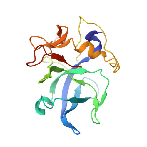
    The EMAPII (endothelial monocyte-activating polypeptide II) domain is a tRNA-binding domain associated with several aminoacyl-tRNA synthetases, which becomes an independent domain with inflammatory cytokine activity upon apoptotic cleavage from the p43 component of the multisynthetase complex. It comprises a domain that is highly homologous to bacterial tRNA-binding proteins (Trbp), followed by an extra domain without homology to known proteins. Trbps, which may represent ancient tRNA chaperones, form dimers and bind one tRNA per dimer. In contrast, EMAPII domains are monomers. Here we report the crystal structure at 1.14 Angstroms of human EMAPII. The structure reveals that the Trbp-like domain, which forms an oligonucleotide-binding (OB) fold, is related by degenerate 2-fold symmetry to the extra-domain. The pseudo-axis coincides with the dyad axis of bacterial TtCsaA, a Trbp whose structure was solved recently. The interdomain interface in EMAPII mimics the intersubunit interface in TtCsaA, and may thus generate a novel OB-fold-based tRNA-binding site. The low sequence homology between the extra domain of EMAPII and either its own OB fold or that of Trbps suggests that dimer mimicry originated from convergent evolution rather than gene duplication.


  • Organizational Affiliation
    • Laboratoire d'Enzymologie et Biochimie Structurales, CNRS, 1 Avenue de la Terrasse, 91198 Gif sur Yvette cedex, France.

Macromolecule Content 

  • Total Structure Weight: 19 kDa 
  • Atom Count: 1,506 
  • Modeled Residue Count: 164 
  • Deposited Residue Count: 171 
  • Unique protein chains: 1

Macromolecules

Find similar proteins by:|  3D Structure
Entity ID: 1
MoleculeChains  Sequence LengthOrganismDetailsImage
ENDOTHELIAL-MONOCYTE ACTIVATING POLYPEPTIDE II171Homo sapiensMutation(s): 0 
UniProt & NIH Common Fund Data Resources
Find proteins for Q12904 (Homo sapiens)
Explore Q12904 
Go to UniProtKB:  Q12904
PHAROS:  Q12904
GTEx:  ENSG00000164022 
Entity Groups
Sequence Clusters30% Identity50% Identity70% Identity90% Identity95% Identity100% Identity
UniProt GroupQ12904
Sequence Annotations
Expand
Reference Sequence

Experimental Data & Validation

Experimental Data

  • Method: X-RAY DIFFRACTION
  • Resolution: 1.50 Å
  • R-Value Free:  0.224 (Depositor) 
  • R-Value Work:  0.221 (Depositor) 
  • R-Value Observed: 0.221 (Depositor) 
Space Group: P 1 21 1
Unit Cell:
Length ( Å )Angle ( ˚ )
a = 35.954α = 90
b = 59.683β = 114.83
c = 37.85γ = 90
Software Package:
Software NamePurpose
MLPHAREphasing
ARP/wARPmodel building
CNSrefinement
DENZOdata reduction
SCALEPACKdata scaling

Structure Validation

View Full Validation Report


Entry History 

Deposition Data

Revision History  (Full details and data files)

  • Version 1.0: 2000-12-06
    Type: Initial release
  • Version 1.1: 2008-04-27
    Changes: Version format compliance
  • Version 1.2: 2011-07-13
    Changes: Version format compliance
  • Version 1.3: 2017-02-08
    Changes: Structure summary
  • Version 1.4: 2024-02-07
    Changes: Data collection, Database references