1FCH | pdb_00001fch

CRYSTAL STRUCTURE OF THE PTS1 COMPLEXED TO THE TPR REGION OF HUMAN PEX5


Experimental Data Snapshot

  • Method: X-RAY DIFFRACTION
  • Resolution: 2.20 Å
  • R-Value Free: 
    0.224 (Depositor), 0.222 (DCC) 
  • R-Value Work: 
    0.194 (Depositor), 0.192 (DCC) 
  • R-Value Observed: 
    0.194 (Depositor) 

wwPDB Validation 3D Report Full Report

Validation slider image for 1FCH

This is version 1.3 of the entry. See complete history

Literature

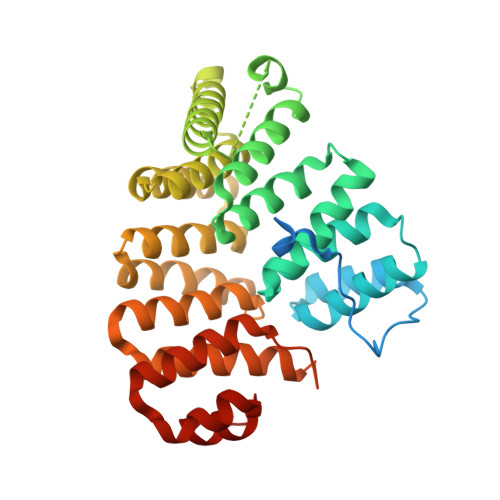
Peroxisomal targeting signal-1 recognition by the TPR domains of human PEX5.

Gatto Jr., G.J.Geisbrecht, B.V.Gould, S.J.Berg, J.M.

(2000) Nat Struct Biol 7: 1091-1095

  • DOI: https://doi.org/10.1038/81930
  • Primary Citation Related Structures: 
    1FCH

  • PubMed Abstract: 

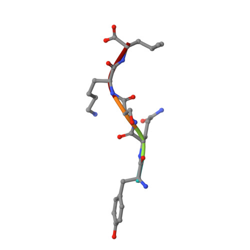
    Many proteins contain targeting signals within their sequences that specify their delivery to particular organelles. The peroxisomal targeting signal-1 (PTS1) is a C-terminal tripeptide that is sufficient to direct proteins into peroxisomes. The PTS1 sequence closely approximates Ser-Lys-Leu-COO-. PEX5, the receptor for PTS1, interacts with the signal via a series of tetratricopeptide repeats (TPRs) within its C-terminal half. Here we report the crystal structure of a fragment of human PEX5 that includes all seven predicted TPR motifs in complex with a pentapeptide containing a PTS1 sequence. Two clusters of three TPRs almost completely surround the peptide, while a hinge region, previously identified as TPR4, forms a distinct structure that enables the two sets of TPRs to form a single binding site. This structure reveals the molecular basis for PTS1 recognition and demonstrates a novel mode of TPR-peptide interaction.


  • Organizational Affiliation
    • Department of Biophysics and Biophysical Chemistry, The Johns Hopkins University School of Medicine, Baltimore, Maryland 21205, USA.

Macromolecule Content 

  • Total Structure Weight: 83.26 kDa 
  • Atom Count: 4,994 
  • Modeled Residue Count: 609 
  • Deposited Residue Count: 746 
  • Unique protein chains: 2

Macromolecules

Find similar proteins by:|  3D Structure
Entity ID: 1
MoleculeChains  Sequence LengthOrganismDetailsImage
PEROXISOMAL TARGETING SIGNAL 1 RECEPTOR
A, B
368Homo sapiensMutation(s): 0 
UniProt & NIH Common Fund Data Resources
Find proteins for P50542 (Homo sapiens)
Explore P50542 
Go to UniProtKB:  P50542
PHAROS:  P50542
GTEx:  ENSG00000139197 
Entity Groups
Sequence Clusters30% Identity50% Identity70% Identity90% Identity95% Identity100% Identity
UniProt GroupP50542
Sequence Annotations
Expand
Reference Sequence
Find similar proteins by:  Sequence   |   3D Structure  
Entity ID: 2
MoleculeChains  Sequence LengthOrganismDetailsImage
PTS1-CONTAINING PEPTIDE
C, D
5N/AMutation(s): 0 

Experimental Data & Validation

Experimental Data

  • Method: X-RAY DIFFRACTION
  • Resolution: 2.20 Å
  • R-Value Free:  0.224 (Depositor), 0.222 (DCC) 
  • R-Value Work:  0.194 (Depositor), 0.192 (DCC) 
  • R-Value Observed: 0.194 (Depositor) 
Space Group: C 1 2 1
Unit Cell:
Length ( Å )Angle ( ˚ )
a = 137.96α = 90
b = 73.51β = 120.78
c = 87.56γ = 90
Software Package:
Software NamePurpose
MLPHAREphasing
CNSrefinement
DENZOdata reduction
SCALEPACKdata scaling

Structure Validation

View Full Validation Report


Entry History 

Deposition Data

Revision History  (Full details and data files)

  • Version 1.0: 2000-12-06
    Type: Initial release
  • Version 1.1: 2008-04-27
    Changes: Version format compliance
  • Version 1.2: 2011-07-13
    Changes: Version format compliance
  • Version 1.3: 2024-02-07
    Changes: Data collection, Database references