1FBY | pdb_00001fby

CRYSTAL STRUCTURE OF THE HUMAN RXR ALPHA LIGAND BINDING DOMAIN BOUND TO 9-CIS RETINOIC ACID


Experimental Data Snapshot

  • Method: X-RAY DIFFRACTION
  • Resolution: 2.25 Å
  • R-Value Free: 
    0.263 (Depositor) 
  • R-Value Work: 
    0.228 (Depositor) 
  • R-Value Observed: 
    0.228 (Depositor) 

wwPDB Validation 3D Report Full Report

Validation slider image for 1FBY

This is version 1.4 of the entry. See complete history

Literature

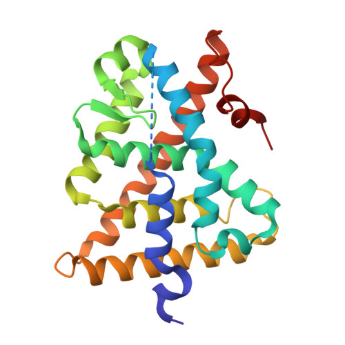
Crystal structure of the human RXRalpha ligand-binding domain bound to its natural ligand: 9-cis retinoic acid.

Egea, P.F.Mitschler, A.Rochel, N.Ruff, M.Chambon, P.Moras, D.

(2000) EMBO J 19: 2592-2601

  • DOI: https://doi.org/10.1093/emboj/19.11.2592
  • Primary Citation Related Structures: 
    1FBY

  • PubMed Abstract: 

    The pleiotropic effects of active retinoids are transduced by their cognate nuclear receptors, retinoid X receptors (RXRs) and retinoic acid receptors (RARs), which act as transcriptional regulators activated by two stereoisomers of retinoic acid (RA): 9-cis RA (9-cRA) and all-trans RA (a-tRA). Among nuclear receptors, RXR occupies a central position and plays a crucial role in many intracellular signalling pathways as a ubiquitous heterodimerization partner with numerous other members of this superfamily. Whereas RARs bind both isomers, RXRs exclusively bind 9-cRA. The crystal structure of the ligand-binding domain (LBD) of human RXRalpha bound to 9-cRA reveals the molecular basis of this ligand selectivity and allows a comparison of both apo and holo forms of the same nuclear receptor. In the crystal, the receptor is monomeric and exhibits a canonical agonist conformation without direct contacts between the ligand and the transactivation helix H12. Comparison with the unliganded RXRalpha LBD structure reveals the molecular mechanisms of ligand-induced conformational changes and allows us to describe at the atomic level how these changes generate the proper protein interface involved in nuclear receptor-coactivator interaction.


  • Organizational Affiliation
    • Institut de Génétique et de Biologie Moléculaire et Cellulaire, CNRS/INSERM/ULP/Collège de France, BP 163-67404 Illkirch Cedex, CU de Strasbourg, France.

Macromolecule Content 

  • Total Structure Weight: 54.11 kDa 
  • Atom Count: 3,625 
  • Modeled Residue Count: 429 
  • Deposited Residue Count: 478 
  • Unique protein chains: 1

Macromolecules

Find similar proteins by:|  3D Structure
Entity ID: 1
MoleculeChains  Sequence LengthOrganismDetailsImage
RETINOIC ACID RECEPTOR RXR-ALPHA
A, B
239Homo sapiensMutation(s): 0 
UniProt & NIH Common Fund Data Resources
Find proteins for P19793 (Homo sapiens)
Explore P19793 
Go to UniProtKB:  P19793
PHAROS:  P19793
GTEx:  ENSG00000186350 
Entity Groups
Sequence Clusters30% Identity50% Identity70% Identity90% Identity95% Identity100% Identity
UniProt GroupP19793
Sequence Annotations
Expand
Reference Sequence

Small Molecules

Ligands 1 Unique
IDChains Name / Formula / InChI Key2D Diagram3D Interactions
9CR

Query on 9CR


Download:Ideal Coordinates CCD File
C [auth A],
D [auth B]
(9cis)-retinoic acid
C20 H28 O2
SHGAZHPCJJPHSC-ZVCIMWCZSA-N
Binding Affinity Annotations 
IDSourceBinding Affinity
9CR BindingDB:  1FBY Ki: min: 3.8, max: 583 (nM) from 12 assay(s)
Kd: min: 1.5, max: 1810 (nM) from 20 assay(s)
IC50: min: 4, max: 82 (nM) from 5 assay(s)
EC50: min: 1.5, max: 316 (nM) from 25 assay(s)

Experimental Data & Validation

Experimental Data

  • Method: X-RAY DIFFRACTION
  • Resolution: 2.25 Å
  • R-Value Free:  0.263 (Depositor) 
  • R-Value Work:  0.228 (Depositor) 
  • R-Value Observed: 0.228 (Depositor) 
Space Group: P 21 21 21
Unit Cell:
Length ( Å )Angle ( ˚ )
a = 66.325α = 90
b = 86.358β = 90
c = 90.527γ = 90
Software Package:
Software NamePurpose
AMoREphasing
CNSrefinement
DENZOdata reduction
SCALEPACKdata scaling

Structure Validation

View Full Validation Report


Entry History 

Deposition Data

Revision History  (Full details and data files)

  • Version 1.0: 2000-07-28
    Type: Initial release
  • Version 1.1: 2008-04-27
    Changes: Version format compliance
  • Version 1.2: 2011-07-13
    Changes: Version format compliance
  • Version 1.3: 2016-11-09
    Changes: Non-polymer description
  • Version 1.4: 2024-02-07
    Changes: Data collection, Database references, Derived calculations