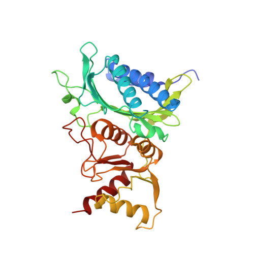
Crystallographic studies of the catalytic mechanism of the neutral form of fructose-1,6-bisphosphatase.
Zhang, Y., Liang, J.Y., Huang, S., Ke, H., Lipscomb, W.N.(1993) Biochemistry 32: 1844-1857
- PubMed: 8382525
- DOI: https://doi.org/10.1021/bi00058a019
- Primary Citation of Related Structures:
1FBC, 1FBD, 1FBE, 1FBF, 1FBG, 1FBH - PubMed Abstract:
The crystal structures of fructose-1,6-bisphosphatase (EC 3.1.3.11) complexed with substrate alone or with substrate analogues in the presence of divalent metal ions have been determined. The substrate analogues, 2,5-anhydro-D-glucitol-1,6-bisphosphate (AhG-1,6-P2) and 2,5-anhydro-D-mannitol-1,6-bisphosphate (AhM-1,6-P2), differ from the alpha and beta anomers of fructose-1,6-bisphosphate (Fru-1,6-P2), respectively, in that the OH on C2 is replaced by a hydrogen atom. Structures have been refined at resolutions of 2.5 to 3.0 A to R factors of 0.172 to 0.195 with root-mean-square deviations of 0.012-0.018 A and 2.7-3.8 degrees from the ideal geometries of bond lengths and bond angles, respectively. In addition, the complex of substrate with the enzyme has been determined in the absence of metal. The electron density at 2.5-A resolution does not distinguish between alpha and beta anomers, which differ for the most part only in the position of the 1-phosphate group and the orientation of the C2-hydroxyl group. The positions of the 6-phosphate and the sugar ring of the substrate analogues are almost identical to those of the respective anomer of the substrate. In the presence of metal ions the positions of the 1-phosphate groups of both alpha and beta analogues differ significantly (0.8-1.0 A) from those of anomers of the substrate in the metal-free complex. Two metal ions (Mn2+ or Zn2+) are located at the enzyme active site of complexes of the alpha analogue AhG-1,6-P2. Metal site 1 is coordinated by the carboxylate groups of Glu-97, Asp-118, and Glu-280 and the 1-phosphate group of substrate analogue, while the metal site 2 is coordinated by the carboxylate groups of Glu-97, Asp-118, the 1-phosphate group of substrate analogue, and the carbonyl oxygen of Leu-120. Both metal sites have a distorted tetrahedral geometry. However, only one metal ion (Mg2+ or Mn2+) is found very near the metal site 1 in the enzyme's active site in complexes of the beta analogue AhM-1,6-P2 or for Mg2+ in the complex of the alpha analogue AhG-1,6-P2. This single metal ion is coordinated by the carboxylate groups of Glu-97, Asp-118, Asp-121, and Glu-280 and the 1-phosphate group of substrate analogue in a distorted square pyramidal geometry.(ABSTRACT TRUNCATED AT 400 WORDS)
Organizational Affiliation:
Gibbs Chemical Laboratory, Harvard University, Cambridge, Massachusetts 02138.


