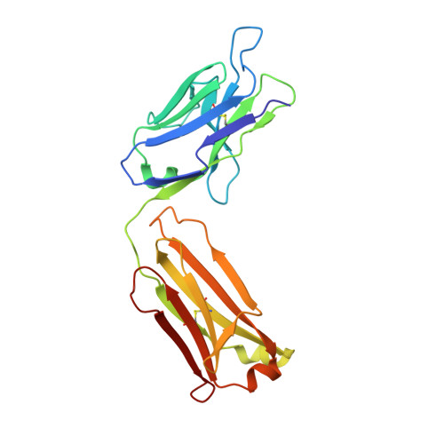
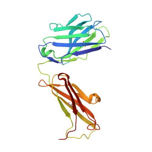
1F8T | pdb_00001f8t

FAB (LNKB-2) OF MONOCLONAL ANTIBODY, CRYSTAL STRUCTURE


Experimental Data Snapshot

  • Method: X-RAY DIFFRACTION
  • Resolution: 2.20 Å
  • R-Value Free: 
    0.221 (Depositor), 0.222 (DCC) 
  • R-Value Work: 
    0.171 (Depositor), 0.176 (DCC) 

wwPDB Validation 3D Report Full Report

Validation slider image for 1F8T

This is version 1.4 of the entry. See complete history. 

Literature

Spatial structure of a Fab-fragment of a monoclonal antibody to human interleukin-2 in two crystalline forms at a resolution of 2.2 and 2.9 angstroms

Fokin, A.V., Afonin, P.V., Mikhailova, I.I.u., Tsygannik, I.N., Mareeva, T.I.u., Nesmeianov, V.A., Pangborn, W., Lee, N., Duax, W., Siszak, E., Pletnev, V.Z.

(2000) Bioorg Khim 26: 571-578

  • Primary Citation Related Structures: 
    1F8T

  • PubMed Abstract: 

    The three-dimensional structure of the antigen-binding fragment of a monoclonal antibody to human interleukin-2 was determined in two crystal forms by the X-ray method of molecular replacement at 2.2 and 2.9 A resolutions. The spatial structure of the protein and the stereochemistry of its antigen-binding site were analyzed.


  • Organizational Affiliation: 
    • Shemyakin-Ovchinnikov Institute of Bioorganic Chemistry, Russian Academy of Sciences, Moscow, Russia.

Macromolecule Content 

  • Total Structure Weight: 48.06 kDa 
  • Atom Count: 3,593 
  • Modeled Residue Count: 439 
  • Deposited Residue Count: 439 
  • Unique protein chains: 2

Macromolecules

Find similar proteins by:|  3D Structure
Entity ID: 1
MoleculeChains  Sequence LengthOrganismDetailsImage
ANTIBODY FAB FRAGMENT (LIGHT CHAIN)A [auth L]219Mus musculusMutation(s): 0 
Entity Groups
Sequence Clusters30% Identity50% Identity70% Identity90% Identity95% Identity100% Identity
Sequence Annotations
Expand
Reference Sequence
Find similar proteins by:|  3D Structure
Entity ID: 2
MoleculeChains  Sequence LengthOrganismDetailsImage
ANTIBODY FAB FRAGMENT (HEAVY CHAIN)B [auth H]220Mus musculusMutation(s): 0 
Entity Groups
Sequence Clusters30% Identity50% Identity70% Identity90% Identity95% Identity100% Identity
Sequence Annotations
Expand
Reference Sequence

Experimental Data & Validation

Experimental Data

  • Method: X-RAY DIFFRACTION
  • Resolution: 2.20 Å
  • R-Value Free:  0.221 (Depositor), 0.222 (DCC) 
  • R-Value Work:  0.171 (Depositor), 0.176 (DCC) 
Space Group: P 21 21 21
Unit Cell:
Length ( Å )Angle ( ˚ )
a = 72.24α = 90
b = 72.01β = 90
c = 86.99γ = 90
Software Package:
Software NamePurpose
DENZOdata reduction
SCALEPACKdata scaling
X-PLORmodel building
CNSrefinement
X-PLORphasing

Structure Validation

View Full Validation Report


Entry History 

Deposition Data

Revision History  (Full details and data files)

  • Version 1.0: 2001-07-05
    Type: Initial release
  • Version 1.1: 2008-04-27
    Changes: Version format compliance
  • Version 1.2: 2011-07-13
    Changes: Version format compliance
  • Version 1.3: 2018-04-04
    Changes: Data collection
  • Version 1.4: 2024-11-20
    Changes: Data collection, Database references, Structure summary