1F7X | pdb_00001f7x

SOLUTION STRUCTURE OF C-TERMINAL DOMAIN ZIPA


Experimental Data Snapshot

  • Method: SOLUTION NMR
  • Conformers Calculated: 100 
  • Conformers Submitted: 30 
  • Selection Criteria: structures with acceptable covalent geometry,structures with favorable non-bond energy,structures with the least restraint violations,structures with the lowest energy 

wwPDB Validation 3D Report Full Report

Validation slider image for 1F7X

This is version 1.4 of the entry. See complete history

Literature

Solution structure of ZipA, a crucial component of Escherichia coli cell division.

Moy, F.J.Glasfeld, E.Mosyak, L.Powers, R.

(2000) Biochemistry 39: 9146-9156

  • DOI: https://doi.org/10.1021/bi0009690
  • Primary Citation Related Structures: 
    1F7W, 1F7X

  • PubMed Abstract: 

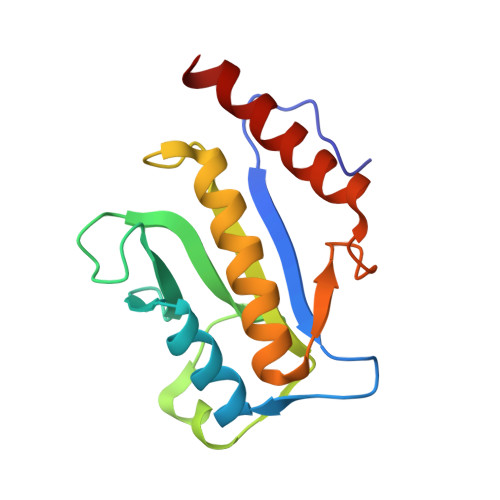
    ZipA, an essential component of cell division in Escherichia coli, interacts with the FtsZ protein at the midcell in one of the initial steps of septum formation. The high-resolution solution structure of the 144-residue C-terminal domain of E. coli ZipA (ZipA(185)(-)(328)) has been determined by multidimensional heteronuclear NMR. A total of 30 structures were calculated by means of hybrid distance geometry-simulated annealing using a total of 2758 experimental NMR restraints. The atomic root means square distribution about the mean coordinate positions for residues 6-142 for the 30 structures is 0.37 +/- 0.04 A for the backbone atoms, 0. 78 +/- 0.05 A for all atoms, and 0.45 +/- 0.04 A for all atoms excluding disordered side chains. The NMR solution structure of ZipA(185)(-)(328) is composed of three alpha-helices and a beta-sheet consisting of six antiparallel beta-strands where the alpha-helices and the beta-sheet form surfaces directly opposite each other. A C-terminal peptide from FtsZ has been shown to bind ZipA(185)(-)(328) in a hydrophobic channel formed by the beta-sheet providing insight into the ZipA-FtsZ interaction. An unexpected similarity between the ZipA(185)(-)(328) fold and the split beta-alpha-beta fold observed in many RNA binding proteins may further our understanding of the critical ZipA-FtsZ interaction.


  • Organizational Affiliation
    • Department of Biological Chemistry, Wyeth Research, Cambridge, Massachusetts 02140, USA.

Macromolecule Content 

  • Total Structure Weight: 16.14 kDa 
  • Atom Count: 1,128 
  • Modeled Residue Count: 144 
  • Deposited Residue Count: 144 
  • Unique protein chains: 1

Macromolecules

Find similar proteins by:|  3D Structure
Entity ID: 1
MoleculeChains  Sequence LengthOrganismDetailsImage
CELL DIVISION PROTEIN ZIPA144Escherichia coliMutation(s): 0 
UniProt
Find proteins for P77173 (Escherichia coli (strain K12))
Explore P77173 
Go to UniProtKB:  P77173
Entity Groups
Sequence Clusters30% Identity50% Identity70% Identity90% Identity95% Identity100% Identity
UniProt GroupP77173
Sequence Annotations
Expand
Reference Sequence

Experimental Data & Validation

Experimental Data

  • Method: SOLUTION NMR
  • Conformers Calculated: 100 
  • Conformers Submitted: 30 
  • Selection Criteria: structures with acceptable covalent geometry,structures with favorable non-bond energy,structures with the least restraint violations,structures with the lowest energy 

Structure Validation

View Full Validation Report


Entry History 

Deposition Data

Revision History  (Full details and data files)

  • Version 1.0: 2001-06-28
    Type: Initial release
  • Version 1.1: 2008-04-27
    Changes: Version format compliance
  • Version 1.2: 2011-07-13
    Changes: Version format compliance
  • Version 1.3: 2022-02-23
    Changes: Data collection, Database references, Derived calculations
  • Version 1.4: 2024-05-22
    Changes: Data collection