1F6E | pdb_00001f6e

CRYSTAL STRUCTURE OF THE A-DNA HEXAMER GGCGM5CC


Experimental Data Snapshot

  • Method: X-RAY DIFFRACTION
  • Resolution: 2.00 Å
  • R-Value Free: 
    0.248 (Depositor) 
  • R-Value Work: 
    0.205 (Depositor), 0.166 (DCC) 
  • R-Value Observed: 
    0.208 (Depositor) 

wwPDB Validation 3D Report Full Report

Validation slider image for 1F6E

This is version 1.3 of the entry. See complete history

Literature

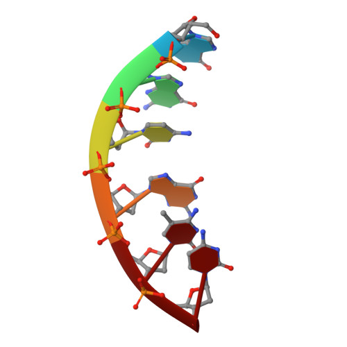
The extended and eccentric E-DNA structure induced by cytosine methylation or bromination.

Vargason, J.M.Eichman, B.F.Ho, P.S.

(2000) Nat Struct Biol 7: 758-761

  • DOI: https://doi.org/10.1038/78985
  • Primary Citation Related Structures: 
    1F69, 1F6C, 1F6E, 1F6I, 1F6J

  • PubMed Abstract: 

    Cytosine methylation or bromination of the DNA sequence d(GGCGCC)2 is shown here to induce a novel extended and eccentric double helix, which we call E-DNA. Like B-DNA, E-DNA has a long helical rise and bases perpendicular to the helix axis. However, the 3'-endo sugar conformation gives the characteristic deep major groove and shallow minor groove of A-DNA. Also, if allowed to crystallize for a period of time longer than that yielding E-DNA, the methylated sequence forms standard A-DNA, suggesting that E-DNA is a kinetically trapped intermediate in the transition to A-DNA. Thus, the structures presented here chart a crystallographic pathway from B-DNA to A-DNA through the E-DNA intermediate in a single sequence. The E-DNA surface is highly accessible to solvent, with waters in the major groove sitting on exposed faces of the stacked nucleotides. We suggest that the geometry of the waters and the stacked base pairs would promote the spontaneous deamination of 5-methylcytosine in the transition mutation of dm5C-dG to dT-dA base pairs.


  • Organizational Affiliation
    • Department of Biochemistry and Biophysics, ALS 2011, Oregon State University, Corvallis, Oregon 97331-7305, USA.

Macromolecule Content 

  • Total Structure Weight: 10.95 kDa 
  • Atom Count: 819 
  • Modeled Residue Count: 36 
  • Deposited Residue Count: 36 
  • Unique nucleic acid chains: 1

Macromolecules

Find similar nucleic acids by:  Sequence
Entity ID: 1
MoleculeChains LengthOrganismImage
DNA (5'-D(*GP*GP*CP*GP*(5CM)P*C)-3')
A, B, C, D, E
A, B, C, D, E, F
6N/A
Sequence Annotations
Expand
Reference Sequence

Experimental Data & Validation

Experimental Data

  • Method: X-RAY DIFFRACTION
  • Resolution: 2.00 Å
  • R-Value Free:  0.248 (Depositor) 
  • R-Value Work:  0.205 (Depositor), 0.166 (DCC) 
  • R-Value Observed: 0.208 (Depositor) 
Space Group: C 2 2 21
Unit Cell:
Length ( Å )Angle ( ˚ )
a = 37.06α = 90
b = 46.727β = 90
c = 110.689γ = 90
Software Package:
Software NamePurpose
AMoREphasing
CNSrefinement
DENZOdata reduction
SCALEPACKdata scaling

Structure Validation

View Full Validation Report


Entry History 

Deposition Data

Revision History  (Full details and data files)

  • Version 1.0: 2000-08-28
    Type: Initial release
  • Version 1.1: 2008-04-27
    Changes: Version format compliance
  • Version 1.2: 2011-07-13
    Changes: Version format compliance
  • Version 1.3: 2024-02-07
    Changes: Data collection, Database references, Derived calculations