1F4R | pdb_00001f4r

CRYSTAL STRUCTURE OF THE HUMAN AAG DNA REPAIR GLYCOSYLASE COMPLEXED WITH 1,N6-ETHENOADENINE-DNA


Experimental Data Snapshot

  • Method: X-RAY DIFFRACTION
  • Resolution: 2.40 Å
  • R-Value Free: 
    0.276 (Depositor) 
  • R-Value Work: 
    0.239 (Depositor) 

wwPDB Validation 3D Report Full Report

Validation slider image for 1F4R

This is version 1.3 of the entry. See complete history

Literature

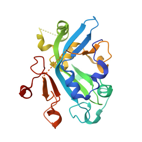
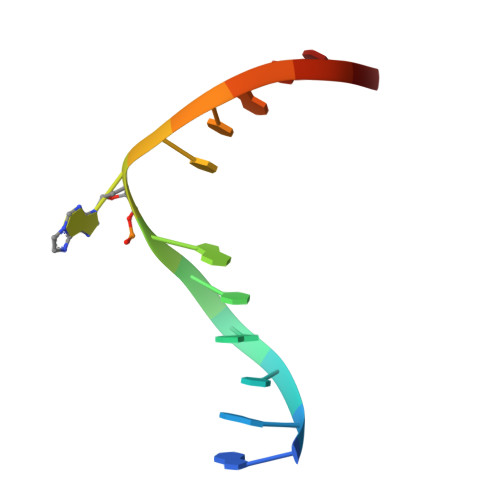
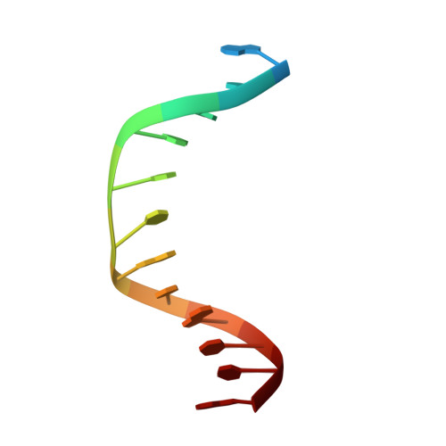
Molecular basis for discriminating between normal and damaged bases by the human alkyladenine glycosylase, AAG.

Lau, A.Y.Wyatt, M.D.Glassner, B.J.Samson, L.D.Ellenberger, T.

(2000) Proc Natl Acad Sci U S A 97: 13573-13578

  • DOI: https://doi.org/10.1073/pnas.97.25.13573
  • Primary Citation Related Structures: 
    1EWN, 1F4R, 1F6O

  • PubMed Abstract: 

    The human 3-methyladenine DNA glycosylase [alkyladenine DNA glycosylase (AAG)] catalyzes the first step of base excision repair by cleaving damaged bases from DNA. Unlike other DNA glycosylases that are specific for a particular type of damaged base, AAG excises a chemically diverse selection of substrate bases damaged by alkylation or deamination. The 2.1-A crystal structure of AAG complexed to DNA containing 1,N(6)-ethenoadenine suggests how modified bases can be distinguished from normal DNA bases in the enzyme active site. Mutational analyses of residues contacting the alkylated base in the crystal structures suggest that the shape of the damaged base, its hydrogen-bonding characteristics, and its aromaticity all contribute to the selective recognition of damage by AAG.


  • Organizational Affiliation
    • Department of Biological Chemistry and Molecular Pharmacology, Harvard Medical School, 240 Longwood Avenue, Boston, MA 02115, USA.

Macromolecule Content 

  • Total Structure Weight: 32.32 kDa 
  • Atom Count: 2,064 
  • Modeled Residue Count: 223 
  • Deposited Residue Count: 245 
  • Unique protein chains: 1
  • Unique nucleic acid chains: 2

Macromolecules


Find similar proteins by:|  3D Structure
Entity ID: 3
MoleculeChains  Sequence LengthOrganismDetailsImage
3-METHYL-ADENINE DNA GLYCOSYLASEC [auth A]219Homo sapiensMutation(s): 0 
EC: 3.2.2.20 (PDB Primary Data), 3.2.2.21 (UniProt)
UniProt & NIH Common Fund Data Resources
Find proteins for P29372 (Homo sapiens)
Explore P29372 
Go to UniProtKB:  P29372
PHAROS:  P29372
GTEx:  ENSG00000103152 
Entity Groups
Sequence Clusters30% Identity50% Identity70% Identity90% Identity95% Identity100% Identity
UniProt GroupP29372
Sequence Annotations
Expand
Reference Sequence
Find similar nucleic acids by:  Sequence
Entity ID: 1
MoleculeChains LengthOrganismImage
DNA (5'-D(*GP*AP*CP*AP*TP*GP*(EDA)P*TP*TP*GP*CP*CP*T)-3')A [auth D]13N/A
Sequence Annotations
Expand
Reference Sequence
Find similar nucleic acids by:  Sequence
Entity ID: 2
MoleculeChains LengthOrganismImage
DNA (5'-D(*GP*GP*CP*AP*AP*TP*CP*AP*TP*GP*TP*CP*A)-3')B [auth E]13N/A
Sequence Annotations
Expand
Reference Sequence

Small Molecules

Ligands 1 Unique
IDChains Name / Formula / InChI Key2D Diagram3D Interactions
NA

Query on NA


Download:Ideal Coordinates CCD File
D [auth A]SODIUM ION
Na
FKNQFGJONOIPTF-UHFFFAOYSA-N

Experimental Data & Validation

Experimental Data

  • Method: X-RAY DIFFRACTION
  • Resolution: 2.40 Å
  • R-Value Free:  0.276 (Depositor) 
  • R-Value Work:  0.239 (Depositor) 
Space Group: P 21 21 21
Unit Cell:
Length ( Å )Angle ( ˚ )
a = 42.1α = 90
b = 57.3β = 90
c = 125.5γ = 90
Software Package:
Software NamePurpose
CNSrefinement
DENZOdata reduction
SCALEPACKdata scaling
CNSphasing

Structure Validation

View Full Validation Report


Entry History 

Deposition Data

Revision History  (Full details and data files)

  • Version 1.0: 2000-12-11
    Type: Initial release
  • Version 1.1: 2008-04-27
    Changes: Version format compliance
  • Version 1.2: 2011-07-13
    Changes: Version format compliance
  • Version 1.3: 2024-02-07
    Changes: Data collection, Database references, Derived calculations