1F3J | pdb_00001f3j

HISTOCOMPATIBILITY ANTIGEN I-AG7


Experimental Data Snapshot

  • Method: X-RAY DIFFRACTION
  • Resolution: 3.10 Å
  • R-Value Free: 
    0.299 (Depositor) 
  • R-Value Work: 
    0.221 (Depositor) 
  • R-Value Observed: 
    0.221 (Depositor) 

Starting Model: experimental
View more details

wwPDB Validation 3D Report Full Report

Validation slider image for 1F3J

This is version 1.7 of the entry. See complete history

Literature

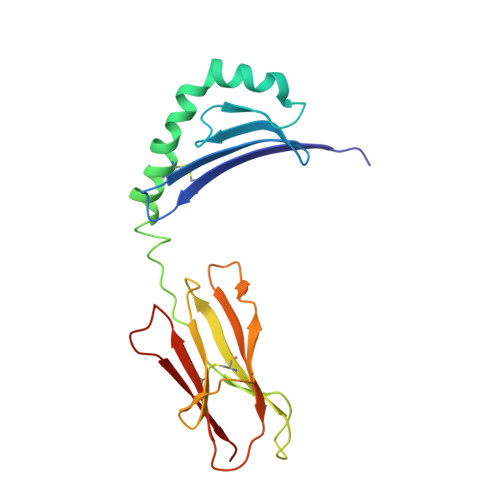
Structural basis of peptide binding and presentation by the type I diabetes-associated MHC class II molecule of NOD mice.

Latek, R.R.Suri, A.Petzold, S.J.Nelson, C.A.Kanagawa, O.Unanue, E.R.Fremont, D.H.

(2000) Immunity 12: 699-710

  • DOI: https://doi.org/10.1016/s1074-7613(00)80220-4
  • Primary Citation Related Structures: 
    1F3J

  • PubMed Abstract: 

    We have determined the crystal structure of I-Ag7, an integral component in murine type I diabetes development. Several features distinguish I-Ag7 from other non-autoimmune-associated MHC class II molecules, including novel peptide and heterodimer pairing interactions. The binding groove of I-Ag7 is unusual at both terminal ends, with a potentially solvent-exposed channel at the base of the P1 pocket and a widened entrance to the P9 pocket. Peptide binding studies with variants of the hen egg lysozyme I-Ag7 epitope HEL(11-25) support a comprehensive structure-based I-Ag7 binding motif. Residues critical for T cell recognition were investigated with a panel of HEL(11-25)-restricted clones, which uncovered P1 anchor-dependent structural variations. These results establish a framework for future experiments directed at understanding the role of I-Ag7 in autoimmunity.


  • Organizational Affiliation
    • Department of Pathology and Immunology, Washington University School of Medicine, St. Louis, Missouri 63110, USA.

Macromolecule Content 

  • Total Structure Weight: 90.62 kDa 
  • Atom Count: 6,464 
  • Modeled Residue Count: 766 
  • Deposited Residue Count: 766 
  • Unique protein chains: 3

Macromolecules

Find similar proteins by:|  3D Structure
Entity ID: 1
MoleculeChains  Sequence LengthOrganismDetailsImage
H-2 CLASS II HISTOCOMPATIBILITY ANTIGEN
A, D
182Mus musculusMutation(s): 0 
UniProt
Find proteins for P04228 (Mus musculus)
Explore P04228 
Go to UniProtKB:  P04228
Entity Groups
Sequence Clusters30% Identity50% Identity70% Identity90% Identity95% Identity100% Identity
UniProt GroupP04228
Glycosylation
Glycosylation Sites: 2
Sequence Annotations
Expand
Reference Sequence
Find similar proteins by:|  3D Structure
Entity ID: 2
MoleculeChains  Sequence LengthOrganismDetailsImage
MHC CLASS II NOD
B, E
187Mus musculusMutation(s): 0 
UniProt
Find proteins for Q31135 (Mus musculus)
Explore Q31135 
Go to UniProtKB:  Q31135
Entity Groups
Sequence Clusters30% Identity50% Identity70% Identity90% Identity95% Identity100% Identity
UniProt GroupQ31135
Glycosylation
Glycosylation Sites: 1
Sequence Annotations
Expand
Reference Sequence
Find similar proteins by:  Sequence   |   3D Structure  
Entity ID: 3
MoleculeChains  Sequence LengthOrganismDetailsImage
LYSOZYME CC [auth P],
F [auth Q]
14Gallus gallusMutation(s): 0 
EC: 3.2.1.17
UniProt
Find proteins for P00698 (Gallus gallus)
Explore P00698 
Go to UniProtKB:  P00698
Entity Groups
Sequence Clusters30% Identity50% Identity70% Identity90% Identity95% Identity100% Identity
UniProt GroupP00698
Sequence Annotations
Expand
Reference Sequence

Experimental Data & Validation

Experimental Data

  • Method: X-RAY DIFFRACTION
  • Resolution: 3.10 Å
  • R-Value Free:  0.299 (Depositor) 
  • R-Value Work:  0.221 (Depositor) 
  • R-Value Observed: 0.221 (Depositor) 
Space Group: P 41 21 2
Unit Cell:
Length ( Å )Angle ( ˚ )
a = 109.54α = 90
b = 109.54β = 90
c = 176.36γ = 90
Software Package:
Software NamePurpose
DENZOdata reduction
SCALEPACKdata scaling
CCP4data reduction
SCALAdata scaling
AMoREphasing
CNSrefinement
CCP4data scaling

Structure Validation

View Full Validation Report


Entry History 

Deposition Data

Revision History  (Full details and data files)

  • Version 1.0: 2000-09-20
    Type: Initial release
  • Version 1.1: 2008-04-27
    Changes: Version format compliance
  • Version 1.2: 2011-07-13
    Changes: Non-polymer description, Version format compliance
  • Version 1.3: 2011-11-16
    Changes: Atomic model
  • Version 1.4: 2017-10-04
    Changes: Refinement description
  • Version 1.5: 2020-07-29
    Type: Remediation
    Reason: Carbohydrate remediation
    Changes: Data collection, Derived calculations, Structure summary
  • Version 1.6: 2023-08-09
    Changes: Data collection, Database references, Refinement description, Structure summary
  • Version 1.7: 2024-10-30
    Changes: Structure summary