1F0O

PVUII ENDONUCLEASE/COGNATE DNA COMPLEX (GLUTARALDEHYDE-CROSSLINKED CRYSTAL) AT PH 7.5 WITH TWO CALCIUM IONS AT EACH ACTIVE SITE


Experimental Data Snapshot

  • Method: X-RAY DIFFRACTION
  • Resolution: 2.50 Å
  • R-Value Free: 0.279 
  • R-Value Work: 0.205 
  • R-Value Observed: 0.279 

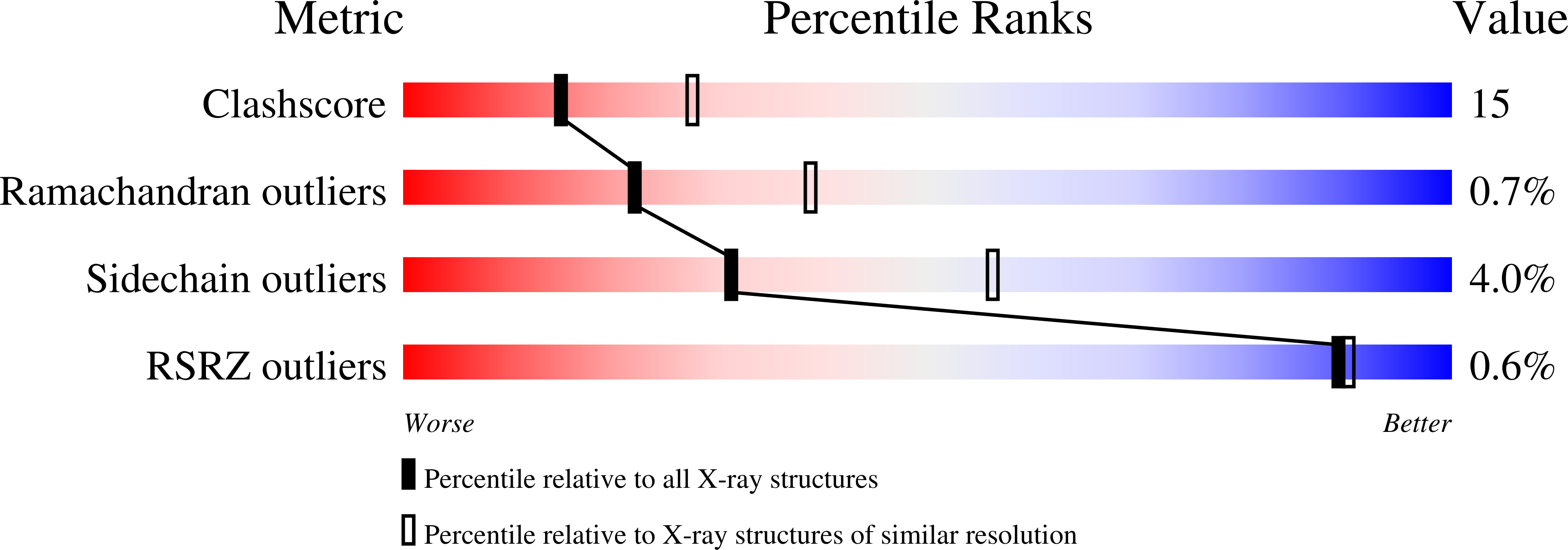
wwPDB Validation   3D Report Full Report


This is version 1.4 of the entry. See complete history


Literature

PvuII endonuclease contains two calcium ions in active sites.

Horton, J.R.Cheng, X.

(2000) J Mol Biol 300: 1049-1056

  • DOI: https://doi.org/10.1006/jmbi.2000.3938
  • Primary Citation of Related Structures:  
    1EYU, 1F0O

  • PubMed Abstract: 

    Restriction endonucleases differ in their use of metal cofactors despite having remarkably similar folds for their catalytic regions. To explore this, we have characterized the interaction of endonuclease PvuII with the catalytically incompetent cation Ca(2+). The structure of a glutaraldehyde-crosslinked crystal of the endonuclease PvuII-DNA complex, determined in the presence of Ca(2+) at a pH of approximately 6.5, supports a two-metal mechanism of DNA cleavage by PvuII. The first Ca(2+) position matches that found in all structurally examined endonucleases, while the second position is similar to that of EcoRV but is distinct from that of BamHI and BglI. The location of the second metal in PvuII, unlike that in BamHI/BglI, permits no direct interaction between the second metal and the O3' oxygen leaving group. However, the interactions between the DNA scissile phosphate and the metals, the first metal and the attacking water, and the attacking water and DNA are the same in PvuII as they are in the two-metal models of BamHI and BglI, but are distinct from the proposed three-metal or the two-metal models of EcoRV.


  • Organizational Affiliation

    Department of Biochemistry, Emory University School of Medicine, 1510 Clifton Road, Atlanta, GA 30322, USA.


Macromolecules

Find similar proteins by:  (by identity cutoff)  |  3D Structure
Entity ID: 2
MoleculeChains Sequence LengthOrganismDetailsImage
TYPE II RESTRICTION ENZYME PVUIIC [auth A],
D [auth B]
157Proteus vulgarisMutation(s): 0 
EC: 3.1.21.4
UniProt
Find proteins for P23657 (Proteus hauseri)
Explore P23657 
Go to UniProtKB:  P23657
Entity Groups  
Sequence Clusters30% Identity50% Identity70% Identity90% Identity95% Identity100% Identity
UniProt GroupP23657
Sequence Annotations
Expand
  • Reference Sequence

Find similar nucleic acids by:  Sequence   |   3D Structure  

Entity ID: 1
MoleculeChains LengthOrganismImage
(5'-D(*TP*GP*AP*CP*CP*AP*GP*CP*TP*GP*GP*TP*C-3')A [auth C],
B [auth D]
13N/A
Sequence Annotations
Expand
  • Reference Sequence
Experimental Data & Validation

Experimental Data

  • Method: X-RAY DIFFRACTION
  • Resolution: 2.50 Å
  • R-Value Free: 0.279 
  • R-Value Work: 0.205 
  • R-Value Observed: 0.279 
  • Space Group: P 21 21 21
Unit Cell:
Length ( Å )Angle ( ˚ )
a = 94.043α = 90
b = 84.702β = 90
c = 45.993γ = 90
Software Package:
Software NamePurpose
X-PLORmodel building
X-PLORrefinement
SCALEPACKdata scaling
X-PLORphasing

Structure Validation

View Full Validation Report


Entry History 

Deposition Data

Revision History  (Full details and data files)

  • Version 1.0: 2000-11-06
    Type: Initial release
  • Version 1.1: 2008-04-27
    Changes: Version format compliance
  • Version 1.2: 2011-07-13
    Changes: Version format compliance
  • Version 1.3: 2017-10-04
    Changes: Refinement description
  • Version 1.4: 2024-02-07
    Changes: Data collection, Database references, Derived calculations