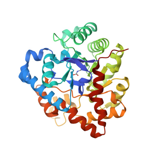
The binding of substrate analogs to phosphotriesterase.
Benning, M.M., Hong, S.B., Raushel, F.M., Holden, H.M.(2000) J Biological Chem 275: 30556-30560
- PubMed: 10871616
- DOI: https://doi.org/10.1074/jbc.M003852200
- Primary Citation of Related Structures:
1EYW, 1EZ2 - PubMed Abstract:
Phosphotriesterase (PTE) from Pseudomonas diminuta catalyzes the detoxification of organophosphates such as the widely utilized insecticide paraoxon and the chemical warfare agent sarin. The three-dimensional structure of the enzyme is known from high resolution x-ray crystallographic analyses. Each subunit of the homodimer folds into a so-called TIM barrel, with eight strands of parallel beta-sheet. The two zinc ions required for activity are positioned at the C-terminal portion of the beta-barrel. Here, we describe the three-dimensional structure of PTE complexed with the inhibitor diisopropyl methyl phosphonate, which serves as a mimic for sarin. Additionally, the structure of the enzyme complexed with triethyl phosphate is also presented. In the case of the PTE-diisopropyl methyl phosphonate complex, the phosphoryl oxygen of the inhibitor coordinates to the more solvent-exposed zinc ion (2.5 A), thereby lending support to the presumed catalytic mechanism involving metal coordination of the substrate. In the PTE-triethyl phosphate complex, the phosphoryl oxygen of the inhibitor is positioned at 3.4 A from the more solvent-exposed zinc ion. The two structures described in this report provide additional molecular understanding for the ability of this remarkable enzyme to hydrolyze such a wide range of organophosphorus substrates.
Organizational Affiliation:
Department of Biochemistry, University of Wisconsin, Madison, USA.


