1EYG | pdb_00001eyg

Crystal structure of chymotryptic fragment of E. coli ssb bound to two 35-mer single strand DNAS


Experimental Data Snapshot

  • Method: X-RAY DIFFRACTION
  • Resolution: 2.80 Å
  • R-Value Free: 
    0.298 (Depositor) 
  • R-Value Work: 
    0.256 (Depositor) 
  • R-Value Observed: 
    0.256 (Depositor) 

wwPDB Validation 3D Report Full Report

Validation slider image for 1EYG

This is version 1.3 of the entry. See complete history

Literature

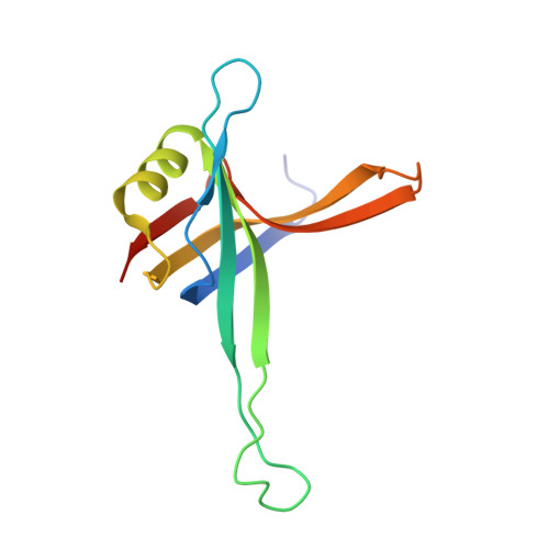
Structure of the DNA binding domain of E. coli SSB bound to ssDNA.

Raghunathan, S.Kozlov, A.G.Lohman, T.M.Waksman, G.

(2000) Nat Struct Biol 7: 648-652

  • DOI: https://doi.org/10.1038/77943
  • Primary Citation Related Structures: 
    1EYG

  • PubMed Abstract: 

    The structure of the homotetrameric DNA binding domain of the single stranded DNA binding protein from Escherichia coli (Eco SSB) bound to two 35-mer single stranded DNAs was determined to a resolution of 2.8 A. This structure describes the vast network of interactions that results in the extensive wrapping of single stranded DNA around the SSB tetramer and suggests a structural basis for its various binding modes.


  • Organizational Affiliation
    • Department of Biochemistry and Molecular Biophysics, Washington University School of Medicine, Campus Box 8231, 660 S. Euclid Ave., Saint Louis, Missouri 63110, USA.

Macromolecule Content 

  • Total Structure Weight: 71.78 kDa 
  • Atom Count: 4,244 
  • Modeled Residue Count: 491 
  • Deposited Residue Count: 534 
  • Unique protein chains: 1
  • Unique nucleic acid chains: 1

Macromolecules


Find similar proteins by:|  3D Structure
Entity ID: 2
MoleculeChains  Sequence LengthOrganismDetailsImage
SINGLE-STRAND DNA-BINDING PROTEINC [auth A],
D [auth B],
E [auth C],
F [auth D]
116Escherichia coliMutation(s): 0 
UniProt
Find proteins for P0AGE0 (Escherichia coli (strain K12))
Explore P0AGE0 
Go to UniProtKB:  P0AGE0
Entity Groups
Sequence Clusters30% Identity50% Identity70% Identity90% Identity95% Identity100% Identity
UniProt GroupP0AGE0
Sequence Annotations
Expand
Reference Sequence
Find similar nucleic acids by:  (by identity cutoff) 
Entity ID: 1
MoleculeChains LengthOrganismImage
SINGLE STRANDED 28-MER OF D(C)A [auth Q],
B [auth R]
35N/A
Sequence Annotations
Expand
Reference Sequence

Experimental Data & Validation

Experimental Data

  • Method: X-RAY DIFFRACTION
  • Resolution: 2.80 Å
  • R-Value Free:  0.298 (Depositor) 
  • R-Value Work:  0.256 (Depositor) 
  • R-Value Observed: 0.256 (Depositor) 
Space Group: C 1 2 1
Unit Cell:
Length ( Å )Angle ( ˚ )
a = 98.688α = 90
b = 71.081β = 91.93
c = 79.16γ = 90
Software Package:
Software NamePurpose
DENZOdata reduction
SCALEPACKdata scaling
SHARPphasing
CNSrefinement

Structure Validation

View Full Validation Report


Entry History 

Deposition Data

Revision History  (Full details and data files)

  • Version 1.0: 2000-08-01
    Type: Initial release
  • Version 1.1: 2008-04-27
    Changes: Version format compliance
  • Version 1.2: 2011-07-13
    Changes: Version format compliance
  • Version 1.3: 2024-02-07
    Changes: Data collection, Database references