1EVT | pdb_00001evt

CRYSTAL STRUCTURE OF FGF1 IN COMPLEX WITH THE EXTRACELLULAR LIGAND BINDING DOMAIN OF FGF RECEPTOR 1 (FGFR1)


Experimental Data Snapshot

  • Method: X-RAY DIFFRACTION
  • Resolution: 2.80 Å
  • R-Value Free: 
    0.300 (Depositor) 
  • R-Value Work: 
    0.249 (Depositor) 
  • R-Value Observed: 
    0.249 (Depositor) 

wwPDB Validation 3D Report Full Report

Validation slider image for 1EVT

This is version 1.3 of the entry. See complete history

Literature

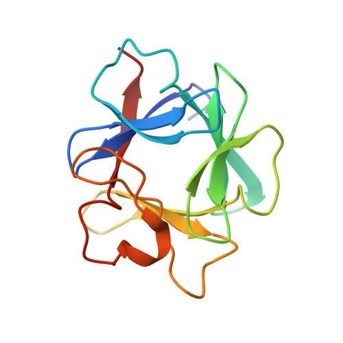
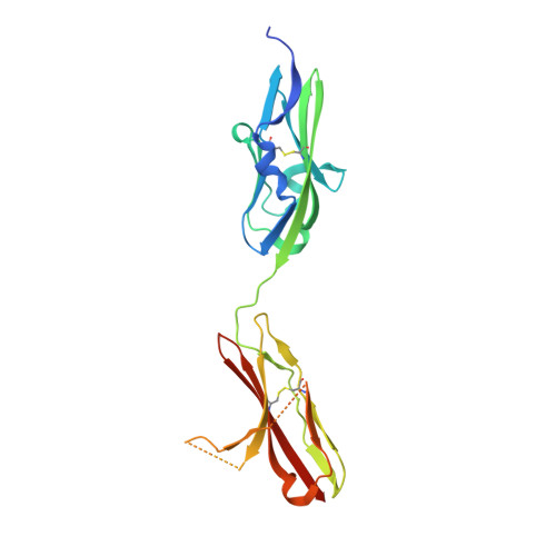
Crystal structures of two FGF-FGFR complexes reveal the determinants of ligand-receptor specificity.

Plotnikov, A.N.Hubbard, S.R.Schlessinger, J.Mohammadi, M.

(2000) Cell 101: 413-424

  • DOI: https://doi.org/10.1016/s0092-8674(00)80851-x
  • Primary Citation Related Structures: 
    1EV2, 1EVT

  • PubMed Abstract: 

    To elucidate the structural determinants governing specificity in fibroblast growth factor (FGF) signaling, we have determined the crystal structures of FGF1 and FGF2 complexed with the ligand binding domains (immunoglobulin-like domains 2 [D2] and 3 [D3]) of FGF receptor 1 (FGFR1) and FGFR2, respectively. Highly conserved FGF-D2 and FGF-linker (between D2-D3) interfaces define a general binding site for all FGF-FGFR complexes. Specificity is achieved through interactions between the N-terminal and central regions of FGFs and two loop regions in D3 that are subject to alternative splicing. These structures provide a molecular basis for FGF1 as a universal FGFR ligand and for modulation of FGF-FGFR specificity through primary sequence variations and alternative splicing.


  • Organizational Affiliation
    • Department of Pharmacology, New York University School of Medicine, New York 10016, USA.

Macromolecule Content 

  • Total Structure Weight: 81.37 kDa 
  • Atom Count: 4,983 
  • Modeled Residue Count: 645 
  • Deposited Residue Count: 718 
  • Unique protein chains: 2

Macromolecules

Find similar proteins by:|  3D Structure
Entity ID: 1
MoleculeChains  Sequence LengthOrganismDetailsImage
PROTEIN (FIBROBLAST GROWTH FACTOR 1)
A, B
134Homo sapiensMutation(s): 0 
UniProt & NIH Common Fund Data Resources
Find proteins for P05230 (Homo sapiens)
Explore P05230 
Go to UniProtKB:  P05230
PHAROS:  P05230
GTEx:  ENSG00000113578 
Entity Groups
Sequence Clusters30% Identity50% Identity70% Identity90% Identity95% Identity100% Identity
UniProt GroupP05230
Sequence Annotations
Expand
Reference Sequence
Find similar proteins by:|  3D Structure
Entity ID: 2
MoleculeChains  Sequence LengthOrganismDetailsImage
PROTEIN (FIBROBLAST GROWTH FACTOR RECEPTOR 1)
C, D
225Homo sapiensMutation(s): 0 
EC: 2.7.10.1
UniProt & NIH Common Fund Data Resources
Find proteins for P11362 (Homo sapiens)
Explore P11362 
Go to UniProtKB:  P11362
PHAROS:  P11362
GTEx:  ENSG00000077782 
Entity Groups
Sequence Clusters30% Identity50% Identity70% Identity90% Identity95% Identity100% Identity
UniProt GroupP11362
Sequence Annotations
Expand
Reference Sequence

Experimental Data & Validation

Experimental Data

  • Method: X-RAY DIFFRACTION
  • Resolution: 2.80 Å
  • R-Value Free:  0.300 (Depositor) 
  • R-Value Work:  0.249 (Depositor) 
  • R-Value Observed: 0.249 (Depositor) 
Space Group: P 1
Unit Cell:
Length ( Å )Angle ( ˚ )
a = 62.553α = 93.4
b = 64.061β = 111.17
c = 64.139γ = 97.18
Software Package:
Software NamePurpose
DENZOdata reduction
SCALEPACKdata scaling
AMoREphasing
CNSrefinement

Structure Validation

View Full Validation Report


Entry History 

Deposition Data

Revision History  (Full details and data files)

  • Version 1.0: 2000-05-31
    Type: Initial release
  • Version 1.1: 2008-04-27
    Changes: Version format compliance
  • Version 1.2: 2011-07-13
    Changes: Version format compliance
  • Version 1.3: 2024-11-13
    Changes: Data collection, Database references, Derived calculations, Structure summary