1EPB | pdb_00001epb

STRUCTURE OF THE EPIDIDYMAL RETINOIC ACID-BINDING PROTEIN AT 2.1 ANGSTROMS RESOLUTION


Experimental Data Snapshot

  • Method: X-RAY DIFFRACTION
  • Resolution: 2.20 Å
  • R-Value Work: 
    0.182 (Depositor) 
  • R-Value Observed: 
    0.182 (Depositor) 

wwPDB Validation 3D Report Full Report

Validation slider image for 1EPB

This is version 1.5 of the entry. See complete history

Literature

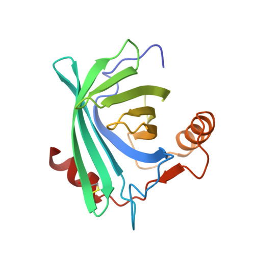
Structure of the epididymal retinoic acid binding protein at 2.1 A resolution.

Newcomer, M.E.

(1993) Structure 1: 7-18

  • DOI: https://doi.org/10.1016/0969-2126(93)90004-z
  • Primary Citation Related Structures: 
    1EPA, 1EPB

  • PubMed Abstract: 

    Androgen-dependent proteins in the lumen of the epididymis are required for sperm maturation. One of these is a retinoic acid binding protein, E-RABP, which binds both all-trans and 9-cis retinoic acid. The other retinoid-binding proteins whose structures are known do not bind 9-cis retinoids. We describe the X-ray structure determination of E-RABP with and without bound ligand. The ligand binds deep in the beta-barrel of the protein, the beta-ionone ring innermost. The binding site, like the ligand, is amphipathic and the deepest part of the cavity is formed by a ring of aromatic amino acids. The isoprene tail of all-trans retinoic acid is bound in a folded conformation which resembles that of the 9-cis isomer. E-RABP achieves high-affinity binding of both all-trans and 9-cis isomers of retinoic acid by forcing the all-trans form to bind in a folded conformation. The RAR family of nuclear receptors for retinoic acid also binds both isomers, and their binding sites may therefore be similar.


  • Organizational Affiliation
    • Department of Biochemistry, Vanderbilt University School of Medicine, Nashville, Tennessee 37232-0146.

Macromolecule Content 

  • Total Structure Weight: 37.1 kDa 
  • Atom Count: 2,545 
  • Modeled Residue Count: 324 
  • Deposited Residue Count: 328 
  • Unique protein chains: 1

Macromolecules

Find similar proteins by:|  3D Structure
Entity ID: 1
MoleculeChains  Sequence LengthOrganismDetailsImage
EPIDIDYMAL RETINOIC ACID-BINDING PROTEIN
A, B
164Rattus norvegicusMutation(s): 0 
Membrane Entity: Yes 
UniProt
Find proteins for P06911 (Rattus norvegicus)
Explore P06911 
Go to UniProtKB:  P06911
Entity Groups
Sequence Clusters30% Identity50% Identity70% Identity90% Identity95% Identity100% Identity
UniProt GroupP06911
Sequence Annotations
Expand
Reference Sequence

Small Molecules

Ligands 1 Unique
IDChains Name / Formula / InChI Key2D Diagram3D Interactions
9CR

Query on 9CR


Download:Ideal Coordinates CCD File
C [auth A],
D [auth B]
(9cis)-retinoic acid
C20 H28 O2
SHGAZHPCJJPHSC-ZVCIMWCZSA-N

Experimental Data & Validation

Experimental Data

  • Method: X-RAY DIFFRACTION
  • Resolution: 2.20 Å
  • R-Value Work:  0.182 (Depositor) 
  • R-Value Observed: 0.182 (Depositor) 
Space Group: P 1 21 1
Unit Cell:
Length ( Å )Angle ( ˚ )
a = 39.8α = 90
b = 58.89β = 109.44
c = 66.65γ = 90
Software Package:
Software NamePurpose
X-PLORmodel building
X-PLORrefinement
X-PLORphasing

Structure Validation

View Full Validation Report


Entry History 

Deposition Data

Revision History  (Full details and data files)

  • Version 1.0: 1994-07-31
    Type: Initial release
  • Version 1.1: 2008-03-24
    Changes: Version format compliance
  • Version 1.2: 2011-07-13
    Changes: Version format compliance
  • Version 1.3: 2016-11-16
    Changes: Non-polymer description
  • Version 1.4: 2017-11-29
    Changes: Derived calculations, Other
  • Version 1.5: 2024-11-20
    Changes: Data collection, Database references, Derived calculations, Structure summary