1EEM | pdb_00001eem

GLUTATHIONE TRANSFERASE FROM HOMO SAPIENS


Experimental Data Snapshot

  • Method: X-RAY DIFFRACTION
  • Resolution: 2.00 Å
  • R-Value Free: 
    0.271 (Depositor), 0.271 (DCC) 
  • R-Value Work: 
    0.219 (Depositor), 0.217 (DCC) 

wwPDB Validation 3D Report Full Report

Validation slider image for 1EEM

Ligand Structure Quality Assessment 


This is version 1.4 of the entry. See complete history

Literature

Identification, characterization, and crystal structure of the Omega class glutathione transferases.

Board, P.G.Coggan, M.Chelvanayagam, G.Easteal, S.Jermiin, L.S.Schulte, G.K.Danley, D.E.Hoth, L.R.Griffor, M.C.Kamath, A.V.Rosner, M.H.Chrunyk, B.A.Perregaux, D.E.Gabel, C.A.Geoghegan, K.F.Pandit, J.

(2000) J Biological Chem 275: 24798-24806

  • DOI: https://doi.org/10.1074/jbc.M001706200
  • Primary Citation Related Structures: 
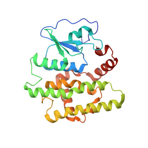
    1EEM

  • PubMed Abstract: 

    A new class of glutathione transferases has been discovered by analysis of the expressed sequence tag data base and sequence alignment. Glutathione S-transferases (GSTs) of the new class, named Omega, exist in several mammalian species and Caenorhabditis elegans. In humans, GSTO 1-1 is expressed in most tissues and exhibits glutathione-dependent thiol transferase and dehydroascorbate reductase activities characteristic of the glutaredoxins. The structure of GSTO 1-1 has been determined at 2.0-A resolution and has a characteristic GST fold (Protein Data Bank entry code ). The Omega class GSTs exhibit an unusual N-terminal extension that abuts the C terminus to form a novel structural unit. Unlike other mammalian GSTs, GSTO 1-1 appears to have an active site cysteine that can form a disulfide bond with glutathione.


  • Organizational Affiliation
    • Molecular Genetics Group and Human Genetics Group, John Curtin School of Medical Research, Australian National University, Canberra, Australian Capital Territory 2601, Australia.

Macromolecule Content 

  • Total Structure Weight: 28.1 kDa 
  • Atom Count: 2,046 
  • Modeled Residue Count: 237 
  • Deposited Residue Count: 241 
  • Unique protein chains: 1

Macromolecules

Find similar proteins by:|  3D Structure
Entity ID: 1
MoleculeChains  Sequence LengthOrganismDetailsImage
GLUTATHIONE-S-TRANSFERASE241Homo sapiensMutation(s): 0 
EC: 1.20.4.2 (UniProt), 1.8.5.1 (UniProt), 2.5.1.18 (UniProt)
UniProt & NIH Common Fund Data Resources
Find proteins for P78417 (Homo sapiens)
Explore P78417 
Go to UniProtKB:  P78417
PHAROS:  P78417
GTEx:  ENSG00000148834 
Entity Groups
Sequence Clusters30% Identity50% Identity70% Identity90% Identity95% Identity100% Identity
UniProt GroupP78417
Sequence Annotations
Expand
Reference Sequence

Biologically Interesting Molecules (External Reference) 

1 Unique

Experimental Data & Validation

Experimental Data

  • Method: X-RAY DIFFRACTION
  • Resolution: 2.00 Å
  • R-Value Free:  0.271 (Depositor), 0.271 (DCC) 
  • R-Value Work:  0.219 (Depositor), 0.217 (DCC) 
Space Group: P 31 2 1
Unit Cell:
Length ( Å )Angle ( ˚ )
a = 57.09α = 90
b = 57.09β = 90
c = 140.28γ = 120
Software Package:
Software NamePurpose
X-PLORmodel building
X-PLORrefinement
DENZOdata reduction
SCALEPACKdata scaling
X-PLORphasing

Structure Validation

View Full Validation Report


Ligand Structure Quality Assessment 


Entry History 

Revision History  (Full details and data files)

  • Version 1.0: 2000-08-11
    Type: Initial release
  • Version 1.1: 2008-04-27
    Changes: Version format compliance
  • Version 1.2: 2011-07-13
    Changes: Version format compliance
  • Version 1.3: 2018-04-04
    Changes: Data collection
  • Version 1.4: 2025-03-26
    Changes: Data collection, Database references, Derived calculations, Structure summary