1EEJ | pdb_00001eej

CRYSTAL STRUCTURE OF THE PROTEIN DISULFIDE BOND ISOMERASE, DSBC, FROM ESCHERICHIA COLI


Experimental Data Snapshot

  • Method: X-RAY DIFFRACTION
  • Resolution: 1.90 Å
  • R-Value Free: 
    0.247 (Depositor), 0.246 (DCC) 
  • R-Value Work: 
    0.214 (Depositor), 0.214 (DCC) 
  • R-Value Observed: 
    0.214 (Depositor) 

wwPDB Validation 3D Report Full Report

Validation slider image for 1EEJ

This is version 1.3 of the entry. See complete history

Literature

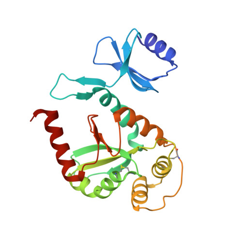
Crystal structure of the protein disulfide bond isomerase, DsbC, from Escherichia coli.

McCarthy, A.A.Haebel, P.W.Torronen, A.Rybin, V.Baker, E.N.Metcalf, P.

(2000) Nat Struct Biol 7: 196-199

  • DOI: https://doi.org/10.1038/73295
  • Primary Citation Related Structures: 
    1EEJ, 1G0T

  • PubMed Abstract: 

    DsbC is one of five Escherichia coli proteins required for disulfide bond formation and is thought to function as a disulfide bond isomerase during oxidative protein folding in the periplasm. DsbC is a 2 x 23 kDa homodimer and has both protein disulfide isomerase and chaperone activity. We report the 1.9 A resolution crystal structure of oxidized DsbC where both Cys-X-X-Cys active sites form disulfide bonds. The molecule consists of separate thioredoxin-like domains joined via hinged linker helices to an N-terminal dimerization domain. The hinges allow relative movement of the active sites, and a broad uncharged cleft between them may be involved in peptide binding and DsbC foldase activities.


  • Organizational Affiliation
    • School of Biological Sciences, Auckland University, Private Bag 92019 Auckland New Zealand.

Macromolecule Content 

  • Total Structure Weight: 47.17 kDa 
  • Atom Count: 3,500 
  • Modeled Residue Count: 432 
  • Deposited Residue Count: 432 
  • Unique protein chains: 1

Macromolecules

Find similar proteins by:|  3D Structure
Entity ID: 1
MoleculeChains  Sequence LengthOrganismDetailsImage
THIOL:DISULFIDE INTERCHANGE PROTEIN
A, B
216Escherichia coliMutation(s): 0 
EC: 5.3.4.1
UniProt
Find proteins for P0AEG6 (Escherichia coli (strain K12))
Explore P0AEG6 
Go to UniProtKB:  P0AEG6
Entity Groups
Sequence Clusters30% Identity50% Identity70% Identity90% Identity95% Identity100% Identity
UniProt GroupP0AEG6
Sequence Annotations
Expand
Reference Sequence

Small Molecules

Ligands 1 Unique
IDChains Name / Formula / InChI Key2D Diagram3D Interactions
MES

Query on MES


Download:Ideal Coordinates CCD File
C [auth B]2-(N-MORPHOLINO)-ETHANESULFONIC ACID
C6 H13 N O4 S
SXGZJKUKBWWHRA-UHFFFAOYSA-N

Experimental Data & Validation

Experimental Data

  • Method: X-RAY DIFFRACTION
  • Resolution: 1.90 Å
  • R-Value Free:  0.247 (Depositor), 0.246 (DCC) 
  • R-Value Work:  0.214 (Depositor), 0.214 (DCC) 
  • R-Value Observed: 0.214 (Depositor) 
Space Group: P 21 21 21
Unit Cell:
Length ( Å )Angle ( ˚ )
a = 59.336α = 90
b = 78.149β = 90
c = 94.489γ = 90
Software Package:
Software NamePurpose
DENZOdata reduction
SCALEPACKdata scaling
SHARPphasing
CNSrefinement

Structure Validation

View Full Validation Report


Entry History 

Deposition Data

Revision History  (Full details and data files)

  • Version 1.0: 2000-08-03
    Type: Initial release
  • Version 1.1: 2008-04-27
    Changes: Version format compliance
  • Version 1.2: 2011-07-13
    Changes: Version format compliance
  • Version 1.3: 2024-10-30
    Changes: Data collection, Database references, Derived calculations, Structure summary