1EDN | pdb_00001edn

HUMAN ENDOTHELIN-1


Experimental Data Snapshot

  • Method: X-RAY DIFFRACTION
  • Resolution: 2.18 Å
  • R-Value Observed: 
    0.199 (Depositor) 

wwPDB Validation 3D Report Full Report

Validation slider image for 1EDN

This is version 1.3 of the entry. See complete history

Literature

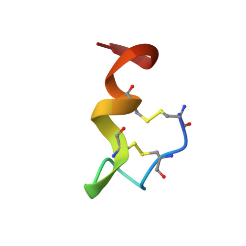
The crystal structure of human endothelin.

Janes, R.W.Peapus, D.H.Wallace, B.A.

(1994) Nat Struct Biol 1: 311-319

  • DOI: https://doi.org/10.1038/nsb0594-311
  • Primary Citation Related Structures: 
    1EDN

  • PubMed Abstract: 

    The three-dimensional structure of the vasoactive polypeptide endothelin, the most potent vasoconstrictor yet identified, has been determined by X-ray crystallography to 2.18 A resolution. This intermediate-sized structure was solved by molecular replacement techniques using a fragment of an NMR-derived model for initial phasing of the data. However, comparisons of the final X-ray structure with the many diverse models derived from NMR data indicate some important differences, especially in the carboxy-terminal region of the molecule: the entire carboxy terminal tail (residues 16-21) is helical in the crystal structure, but not in any of the NMR structures. This may be a functionally significant difference as this region is crucial for receptor binding and vasoactivity.


  • Organizational Affiliation
    • Department of Crystallography, Birkbeck College, University of London, UK.

Macromolecule Content 

  • Total Structure Weight: 2.5 kDa 
  • Atom Count: 171 
  • Modeled Residue Count: 21 
  • Deposited Residue Count: 21 
  • Unique protein chains: 1

Macromolecules

Find similar proteins by:  Sequence   |   3D Structure  
Entity ID: 1
MoleculeChains  Sequence LengthOrganismDetailsImage
ENDOTHELIN-121Homo sapiensMutation(s): 0 
Membrane Entity: Yes 
UniProt & NIH Common Fund Data Resources
Find proteins for P05305 (Homo sapiens)
Explore P05305 
Go to UniProtKB:  P05305
PHAROS:  P05305
GTEx:  ENSG00000078401 
Entity Groups
Sequence Clusters30% Identity50% Identity70% Identity90% Identity95% Identity100% Identity
UniProt GroupP05305
Sequence Annotations
Expand
Reference Sequence

Experimental Data & Validation

Experimental Data

  • Method: X-RAY DIFFRACTION
  • Resolution: 2.18 Å
  • R-Value Observed: 0.199 (Depositor) 
Space Group: P 61 2 2
Unit Cell:
Length ( Å )Angle ( ˚ )
a = 33.35α = 90
b = 33.35β = 90
c = 59.28γ = 120
Software Package:
Software NamePurpose
MOSFLMdata reduction
X-PLORmodel building
X-PLORrefinement
X-PLORphasing

Structure Validation

View Full Validation Report


Entry History 

Deposition Data

Revision History  (Full details and data files)

  • Version 1.0: 1995-10-15
    Type: Initial release
  • Version 1.1: 2008-03-24
    Changes: Version format compliance
  • Version 1.2: 2011-07-13
    Changes: Version format compliance
  • Version 1.3: 2024-10-23
    Changes: Data collection, Database references, Structure summary