1EAP | pdb_00001eap

CRYSTAL STRUCTURE OF A CATALYTIC ANTIBODY WITH A SERINE PROTEASE ACTIVE SITE


Experimental Data Snapshot

  • Method: X-RAY DIFFRACTION
  • Resolution: 2.40 Å
  • R-Value Work: 
    0.186 (Depositor) 
  • R-Value Observed: 
    0.186 (Depositor) 

wwPDB Validation 3D Report Full Report

Validation slider image for 1EAP

This is version 1.4 of the entry. See complete history

Literature

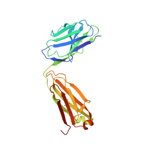
Crystal structure of a catalytic antibody with a serine protease active site.

Zhou, G.W.Guo, J.Huang, W.Scanlan, T.S.Fletterick, R.J.

(1994) Science 265: 1059-1064

  • DOI: https://doi.org/10.1126/science.8066444
  • Primary Citation Related Structures: 
    1EAP

  • PubMed Abstract: 

    The three-dimensional structure of an unusually active hydrolytic antibody with a phosphonate transition state analog (hapten) bound to the active site has been solved to 2.5 A resolution. The antibody (17E8) catalyzes the hydrolysis of norleucine and methionine phenyl esters and is selective for amino acid esters that have the natural alpha-carbon L configuration. A plot of the pH-dependence of the antibody-catalyzed reaction is bell-shaped with an activity maximum at pH 9.5; experiments on mechanism lend support to the formation of a covalent acyl-antibody intermediate. The structural and kinetic data are complementary and support a hydrolytic mechanism for the antibody that is remarkably similar to that of the serine proteases. The antibody active site contains a Ser-His dyad structure proximal to the phosphorous atom of the bound hapten that resembles two of the three components of the Ser-His-Asp catalytic triad of serine proteases. The antibody active site also contains a Lys residue to stabilize oxyanion formation, and a hydrophobic binding pocket for specific substrate recognition of norleucine and methionine side chains. The structure identifies active site residues that mediate catalysis and suggests specific mutations that may improve the catalytic efficiency of the antibody. This high resolution structure of a catalytic antibody-hapten complex shows that antibodies can converge on active site structures that have arisen through natural enzyme evolution.


  • Organizational Affiliation
    • Department of Biochemistry and Biophysics, University of California, San Francisco 94143-0448.

Macromolecule Content 

  • Total Structure Weight: 46.76 kDa 
  • Atom Count: 3,287 
  • Modeled Residue Count: 429 
  • Deposited Residue Count: 429 
  • Unique protein chains: 2

Macromolecules

Find similar proteins by:|  3D Structure
Entity ID: 1
MoleculeChains  Sequence LengthOrganismDetailsImage
IGG2B-KAPPA 17E8 FAB (LIGHT CHAIN)213Mus musculusMutation(s): 0 
Find similar proteins by:|  3D Structure
Entity ID: 2
MoleculeChains  Sequence LengthOrganismDetailsImage
IGG2B-KAPPA 17E8 FAB (HEAVY CHAIN)216Mus musculusMutation(s): 0 

Small Molecules

Ligands 1 Unique
IDChains Name / Formula / InChI Key2D Diagram3D Interactions
HEP

Query on HEP


Download:Ideal Coordinates CCD File
C [auth B]PHENYL[1-(N-SUCCINYLAMINO)PENTYL]PHOSPHONATE
C15 H22 N O6 P
FJQWWGCHPFSERW-CQSZACIVSA-N

Experimental Data & Validation

Experimental Data

  • Method: X-RAY DIFFRACTION
  • Resolution: 2.40 Å
  • R-Value Work:  0.186 (Depositor) 
  • R-Value Observed: 0.186 (Depositor) 
Space Group: P 1 21 1
Unit Cell:
Length ( Å )Angle ( ˚ )
a = 41.41α = 90
b = 78.06β = 94.94
c = 81.94γ = 90
Software Package:
Software NamePurpose
X-PLORmodel building
X-PLORrefinement
X-PLORphasing

Structure Validation

View Full Validation Report


Entry History 

Deposition Data

Revision History  (Full details and data files)

  • Version 1.0: 1994-12-20
    Type: Initial release
  • Version 1.1: 2008-03-24
    Changes: Version format compliance
  • Version 1.2: 2011-07-13
    Changes: Version format compliance
  • Version 1.3: 2024-06-05
    Changes: Data collection, Database references, Derived calculations, Other
  • Version 1.4: 2024-10-09
    Changes: Structure summary