1E9W | pdb_00001e9w

Structure of the macrocycle thiostrepton solved using the anomalous dispersive contribution from sulfur


Experimental Data Snapshot

  • Method: X-RAY DIFFRACTION
  • Resolution: 1.02 Å
  • R-Value Free: 
    0.142 (Depositor), 0.149 (DCC) 
  • R-Value Work: 
    0.118 (DCC) 
  • R-Value Observed: 
    0.112 (Depositor) 

Starting Model: other
View more details

wwPDB Validation 3D Report Full Report

Validation slider image for 1E9W

This is version 2.2 of the entry. See complete history

Literature

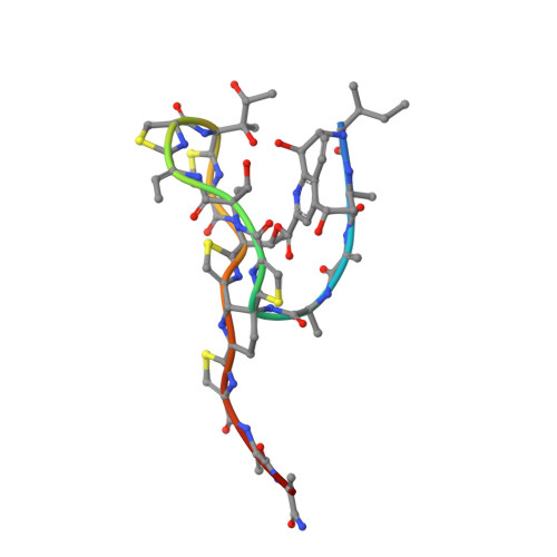
Structure of the Macrocycle Thiostrepton Solved Using the Anomalous Dispersive Contribution from Sulfur

Bond, C.S.Shaw, M.P.Alphey, M.S.Hunter, W.N.

(2001) Acta Crystallogr D Biol Crystallogr 57: 755

  • DOI: https://doi.org/10.1107/s0907444901003134
  • Primary Citation Related Structures: 
    1E9W

  • PubMed Abstract: 

    The structure of a tetragonal crystal form of thiostrepton has been solved using the anomalous dispersive effects of five S atoms from high-redundancy data collected to 1.33 A resolution at the Cu Kalpha wavelength. Data measured to 1.02 A resolution with a synchrotron source were used for refinement. Details of the molecular structure, intramolecular and intermolecular interactions are given.


  • Organizational Affiliation
    • The Wellcome Trust Biocentre, University of Dundee, Dundee DD1 5EH, Scotland, UK.

Macromolecule Content 

  • Total Structure Weight: 1.81 kDa 
  • Atom Count: 118 
  • Modeled Residue Count: 19 
  • Deposited Residue Count: 19 
  • Unique protein chains: 1

Macromolecules

Find similar proteins by:  Sequence   |   3D Structure  
Entity ID: 1
MoleculeChains  Sequence LengthOrganismDetailsImage
THIOSTREPTON19Streptomyces azureusMutation(s): 0 
UniProt
Find proteins for P0C8P8 (Streptomyces azureus)
Explore P0C8P8 
Go to UniProtKB:  P0C8P8
Entity Groups
Sequence Clusters30% Identity50% Identity70% Identity90% Identity95% Identity100% Identity
UniProt GroupP0C8P8
Sequence Annotations
Expand
Reference Sequence

Small Molecules

Modified Residues  5 Unique
IDChains TypeFormula2D DiagramParent
BB9
Query on BB9
A
PEPTIDE LINKINGC3 H5 N O2 SCYS
DBU
Query on DBU
A
PEPTIDE LINKINGC4 H7 N O2THR
DCY
Query on DCY
A
D-PEPTIDE LINKINGC3 H7 N O2 S

--

DHA
Query on DHA
A
PEPTIDE LINKINGC3 H5 N O2SER
MH6
Query on MH6
A
PEPTIDE LINKINGC3 H5 N O3SER
TS9
Query on TS9
A
L-PEPTIDE LINKINGC6 H13 N O4ILE

Biologically Interesting Molecules (External Reference) 

1 Unique

Experimental Data & Validation

Experimental Data

  • Method: X-RAY DIFFRACTION
  • Resolution: 1.02 Å
  • R-Value Free:  0.142 (Depositor), 0.149 (DCC) 
  • R-Value Work:  0.118 (DCC) 
  • R-Value Observed: 0.112 (Depositor) 
Space Group: P 43 21 2
Unit Cell:
Length ( Å )Angle ( ˚ )
a = 26.577α = 90
b = 26.577β = 90
c = 27.436γ = 90
Software Package:
Software NamePurpose
SHELXL-97refinement
DENZOdata reduction
SCALEPACKdata scaling
SHELXphasing

Structure Validation

View Full Validation Report


Entry History 

Deposition Data

Revision History  (Full details and data files)

  • Version 1.0: 2001-03-23
    Type: Initial release
  • Version 1.1: 2011-07-13
    Changes: Atomic model, Database references, Derived calculations, Structure summary, Version format compliance
  • Version 1.2: 2012-11-30
    Changes: Other
  • Version 2.0: 2019-04-24
    Changes: Data collection, Derived calculations, Other, Polymer sequence
  • Version 2.1: 2019-05-22
    Changes: Data collection, Refinement description
  • Version 2.2: 2024-05-01
    Changes: Data collection, Database references, Derived calculations, Other, Refinement description