1E6U | pdb_00001e6u

GDP 4-keto-6-deoxy-D-mannose epimerase reductase


Experimental Data Snapshot

  • Method: X-RAY DIFFRACTION
  • Resolution: 1.45 Å
  • R-Value Free: 
    0.170 (Depositor) 
  • R-Value Work: 
    0.135 (Depositor) 

Starting Model: experimental
View more details

wwPDB Validation 3D Report Full Report

Validation slider image for 1E6U

This is version 1.5 of the entry. See complete history

Literature

Probing the Catalytic Mechanism of Gdp-4-Keto-6-Deoxy-D-Mannose Epimerase/Reductase by Kinetic and Crystallographic Characterization of Site-Specific Mutants

Rosano, C.Bisso, A.Izzo, G.Tonetti, M.Sturla, L.De Flora, A.Bolognesi, M.

(2000) J Mol Biology 303: 77

  • DOI: https://doi.org/10.1006/jmbi.2000.4106
  • Primary Citation Related Structures: 
    1E6U, 1E7Q, 1E7R, 1E7S

  • PubMed Abstract: 

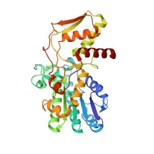
    GDP-4-keto-6-deoxy-d-mannose epimerase/reductase is a bifunctional enzyme responsible for the last step in the biosynthesis of GDP-l-fucose, the substrate of fucosyl transferases. Several cell-surface antigens, including the leukocyte Lewis system and cell-surface antigens in pathogenic bacteria, depend on the availability of GDP-l-fucose for their expression. Therefore, the enzyme is a potential target for therapy in pathological states depending on selectin-mediated cell-to-cell interactions. Previous crystallographic investigations have shown that GDP-4-keto-6-deoxy-d-mannose epimerase/reductase belongs to the short-chain dehydrogenase/reductase protein homology family. The enzyme active-site region is at the interface of an N-terminal NADPH-binding domain and a C-terminal domain, held to bind the substrate. The design, expression and functional characterization of seven site-specific mutant forms of GDP-4-keto-6-deoxy-d-mannose epimerase/reductase are reported here. In parallel, the crystal structures of the native holoenzyme and of three mutants (Ser107Ala, Tyr136Glu and Lys140Arg) have been investigated and refined at 1. 45-1.60 A resolution, based on synchrotron data (R-factors range between 12.6 % and 13.9 %). The refined protein models show that besides the active-site residues Ser107, Tyr136 and Lys140, whose mutations impair the overall enzymatic activity and may affect the coenzyme binding mode, side-chains capable of proton exchange, located around the expected substrate (GDP-4-keto-6-deoxy-d-mannose) binding pocket, are selectively required during the epimerization and reduction steps. Among these, Cys109 and His179 may play a primary role in proton exchange between the enzyme and the epimerization catalytic intermediates. Finally, the additional role of mutated active-site residues involved in substrate recognition and in enzyme stability has been analyzed.


  • Organizational Affiliation
    • Department of Physics-INFM and Advanced Biotechnology Center-IST, University of Genova, Largo Rosanna Benzi 10, Genova, I-16132, Italy.

Macromolecule Content 

  • Total Structure Weight: 37.58 kDa 
  • Atom Count: 2,974 
  • Modeled Residue Count: 315 
  • Deposited Residue Count: 321 
  • Unique protein chains: 1

Macromolecules

Find similar proteins by:|  3D Structure
Entity ID: 1
MoleculeChains  Sequence LengthOrganismDetailsImage
GDP-FUCOSE SYNTHETASE321Escherichia coliMutation(s): 0 
EC: 5.1.3 (PDB Primary Data), 1.1.1.271 (UniProt)
UniProt
Find proteins for P32055 (Escherichia coli (strain K12))
Explore P32055 
Go to UniProtKB:  P32055
Entity Groups
Sequence Clusters30% Identity50% Identity70% Identity90% Identity95% Identity100% Identity
UniProt GroupP32055
Sequence Annotations
Expand
Reference Sequence

Small Molecules

Ligands 4 Unique
IDChains Name / Formula / InChI Key2D Diagram3D Interactions
NAP

Query on NAP


Download:Ideal Coordinates CCD File
B [auth A]NADP NICOTINAMIDE-ADENINE-DINUCLEOTIDE PHOSPHATE
C21 H28 N7 O17 P3
XJLXINKUBYWONI-NNYOXOHSSA-N
UVW

Query on UVW


Download:Ideal Coordinates CCD File
C [auth A]ACETYLPHOSPHATE
C2 H5 O5 P
LIPOUNRJVLNBCD-UHFFFAOYSA-N
TRS

Query on TRS


Download:Ideal Coordinates CCD File
H [auth A]2-AMINO-2-HYDROXYMETHYL-PROPANE-1,3-DIOL
C4 H12 N O3
LENZDBCJOHFCAS-UHFFFAOYSA-O
SO4

Query on SO4


Download:Ideal Coordinates CCD File
D [auth A],
E [auth A],
F [auth A],
G [auth A]
SULFATE ION
O4 S
QAOWNCQODCNURD-UHFFFAOYSA-L

Experimental Data & Validation

Experimental Data

  • Method: X-RAY DIFFRACTION
  • Resolution: 1.45 Å
  • R-Value Free:  0.170 (Depositor) 
  • R-Value Work:  0.135 (Depositor) 
Space Group: P 32 2 1
Unit Cell:
Length ( Å )Angle ( ˚ )
a = 102.8α = 90
b = 102.8β = 90
c = 74.9γ = 120
Software Package:
Software NamePurpose
REFMACrefinement
DENZOdata reduction
SCALEPACKdata scaling

Structure Validation

View Full Validation Report


Entry History 

Deposition Data

Revision History  (Full details and data files)

  • Version 1.0: 2000-10-22
    Type: Initial release
  • Version 1.1: 2011-05-08
    Changes: Version format compliance
  • Version 1.2: 2011-07-13
    Changes: Version format compliance
  • Version 1.3: 2019-05-08
    Changes: Advisory, Data collection, Derived calculations, Experimental preparation, Other
  • Version 1.4: 2023-12-13
    Changes: Advisory, Data collection, Database references, Derived calculations, Refinement description
  • Version 1.5: 2025-10-01
    Changes: Advisory, Derived calculations, Structure summary