1E4E | pdb_00001e4e

D-alanyl-D-lacate ligase


Experimental Data Snapshot

  • Method: X-RAY DIFFRACTION
  • Resolution: 2.50 Å
  • R-Value Free: 
    0.257 (Depositor) 
  • R-Value Work: 
    0.183 (Depositor), 0.178 (DCC) 

Starting Model: experimental
View more details

wwPDB Validation 3D Report Full Report

Validation slider image for 1E4E

Ligand Structure Quality Assessment 


This is version 1.5 of the entry. See complete history

Literature

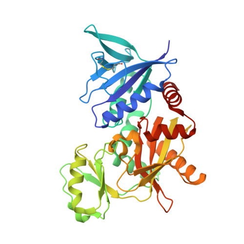
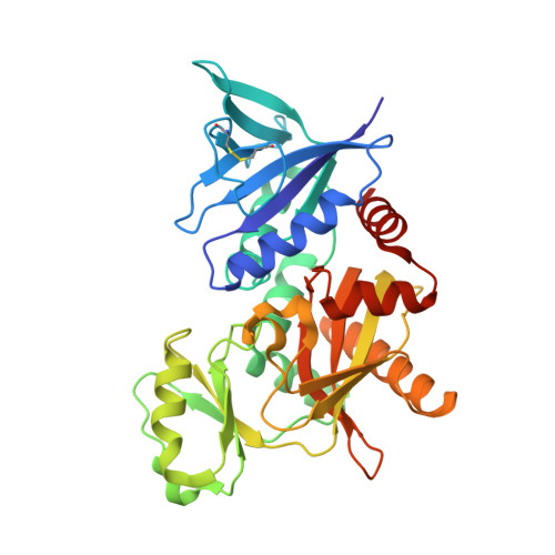
The molecular basis of vancomycin resistance in clinically relevant Enterococci: crystal structure of D-alanyl-D-lactate ligase (VanA).

Roper, D.I.Huyton, T.Vagin, A.Dodson, G.

(2000) Proc Natl Acad Sci U S A 97: 8921-8925

  • DOI: https://doi.org/10.1073/pnas.150116497
  • Primary Citation Related Structures: 
    1E4E

  • PubMed Abstract: 

    d-alanine-d-lactate ligase from Enterococcus faecium BM4147 is directly responsible for the biosynthesis of alternate cell-wall precursors in bacteria, which are resistant to the glycopeptide antibiotic vancomycin. The crystal structure has been determined with data extending to 2.5-A resolution. This structure shows that the active site has unexpected interactions and is distinct from previous models for d-alanyl-d-lactate ligase mechanistic studies. It appears that the preference of the enzyme for lactate as a ligand over d-alanine could be mediated by electrostatic effects and/or a hydrogen-bonding network, which principally involve His-244. The structure of d-alanyl-d-lactate ligase provides a revised interpretation of the molecular events that lead to vancomycin resistance.


  • Organizational Affiliation
    • York Structural Biology Laboratory, Department of Chemistry, The University of York, Heslington, York YO10 5DD, United Kingdom. roper@ysbl.york.ac.uk

Macromolecule Content 

  • Total Structure Weight: 78.27 kDa 
  • Atom Count: 5,750 
  • Modeled Residue Count: 681 
  • Deposited Residue Count: 686 
  • Unique protein chains: 2

Macromolecules

Find similar proteins by:|  3D Structure
Entity ID: 1
MoleculeChains  Sequence LengthOrganismDetailsImage
VANCOMYCIN/TEICOPLANIN A-TYPE RESISTANCE PROTEIN VANA343Enterococcus faeciumMutation(s): 0 
Gene Names: VANA
EC: 6.1.2.1
UniProt
Find proteins for P25051 (Enterococcus faecium)
Explore P25051 
Go to UniProtKB:  P25051
Entity Groups
Sequence Clusters30% Identity50% Identity70% Identity90% Identity95% Identity100% Identity
UniProt GroupP25051
Sequence Annotations
Expand
Reference Sequence
Find similar proteins by:|  3D Structure
Entity ID: 2
MoleculeChains  Sequence LengthOrganismDetailsImage
VANCOMYCIN/TEICOPLANIN A-TYPE RESISTANCE PROTEIN VANA343Enterococcus faeciumMutation(s): 0 
Gene Names: VANA
EC: 6.1.2.1
UniProt
Find proteins for P25051 (Enterococcus faecium)
Explore P25051 
Go to UniProtKB:  P25051
Entity Groups
Sequence Clusters30% Identity50% Identity70% Identity90% Identity95% Identity100% Identity
UniProt GroupP25051
Sequence Annotations
Expand
Reference Sequence

Small Molecules

Ligands 5 Unique
IDChains Name / Formula / InChI Key2D Diagram3D Interactions
ADP

Query on ADP


Download:Ideal Coordinates CCD File
C [auth A],
Q [auth B]
ADENOSINE-5'-DIPHOSPHATE
C10 H15 N5 O10 P2
XTWYTFMLZFPYCI-KQYNXXCUSA-N
PHY

Query on PHY


Download:Ideal Coordinates CCD File
D [auth A],
R [auth B]
1(S)-AMINOETHYL-(2-CARBOXYPROPYL)PHOSPHORYL-PHOSPHINIC ACID
C6 H15 N O7 P2
BAIYWTZQRMCJBV-DKDXWZAISA-N
SO4

Query on SO4


Download:Ideal Coordinates CCD File
G [auth A],
H [auth A],
U [auth B]
SULFATE ION
O4 S
QAOWNCQODCNURD-UHFFFAOYSA-L
GOL

Query on GOL


Download:Ideal Coordinates CCD File
AA [auth B]
I [auth A]
J [auth A]
K [auth A]
L [auth A]
AA [auth B],
I [auth A],
J [auth A],
K [auth A],
L [auth A],
M [auth A],
N [auth A],
O [auth A],
P [auth A],
V [auth B],
W [auth B],
X [auth B],
Y [auth B],
Z [auth B]
GLYCEROL
C3 H8 O3
PEDCQBHIVMGVHV-UHFFFAOYSA-N
MG

Query on MG


Download:Ideal Coordinates CCD File
E [auth A],
F [auth A],
S [auth B],
T [auth B]
MAGNESIUM ION
Mg
JLVVSXFLKOJNIY-UHFFFAOYSA-N

Experimental Data & Validation

Experimental Data

  • Method: X-RAY DIFFRACTION
  • Resolution: 2.50 Å
  • R-Value Free:  0.257 (Depositor) 
  • R-Value Work:  0.183 (Depositor), 0.178 (DCC) 
Space Group: C 2 2 21
Unit Cell:
Length ( Å )Angle ( ˚ )
a = 123.204α = 90
b = 225.359β = 90
c = 72.439γ = 90
Software Package:
Software NamePurpose
REFMACrefinement
SCALEPACKdata scaling
MOLREPphasing

Structure Validation

View Full Validation Report


Ligand Structure Quality Assessment 


Entry History 

Deposition Data

  • Released Date: 2001-06-28 
  • Deposition Author(s): Roper, D.I.

Revision History  (Full details and data files)

  • Version 1.0: 2001-06-28
    Type: Initial release
  • Version 1.1: 2011-09-21
    Changes: Atomic model, Data collection, Derived calculations, Non-polymer description, Other, Source and taxonomy, Structure summary, Version format compliance
  • Version 1.2: 2017-09-13
    Changes: Advisory, Database references
  • Version 1.3: 2019-10-09
    Changes: Data collection, Experimental preparation, Other
  • Version 1.4: 2023-12-13
    Changes: Advisory, Data collection, Database references, Derived calculations, Refinement description
  • Version 1.5: 2024-10-09
    Changes: Structure summary