1E38 | pdb_00001e38

PORCINE PANCREATIC ELASTASE COMPLEXED WITH (3S, 4S)N-PARA-NITROBENZENESULPHONYL -3-ETHYL-4-(CARBOXYLIC ACID)PYRROLIDIN-2-ONE SOAKED IN PH 9 BUFFER FOR 2 MINUTES


Experimental Data Snapshot

  • Method: X-RAY DIFFRACTION
  • Resolution: 1.70 Å
  • R-Value Free: 
    0.220 (Depositor) 
  • R-Value Work: 
    0.183 (Depositor) 

Starting Model: experimental
View more details

wwPDB Validation 3D Report Full Report

Validation slider image for 1E38

This is version 1.4 of the entry. See complete history

Literature

'Ph-Jump' Crystallographic Analyses of Gamma-Lactam-Porcine Pancreatic Elastase Complexes

Wright, P.A.Wilmouth, R.C.Clifton, I.J.Schofield, C.J.

(2000) Biochem J 351: 335

  • Primary Citation Related Structures: 
    1E34, 1E35, 1E36, 1E37, 1E38

  • PubMed Abstract: 

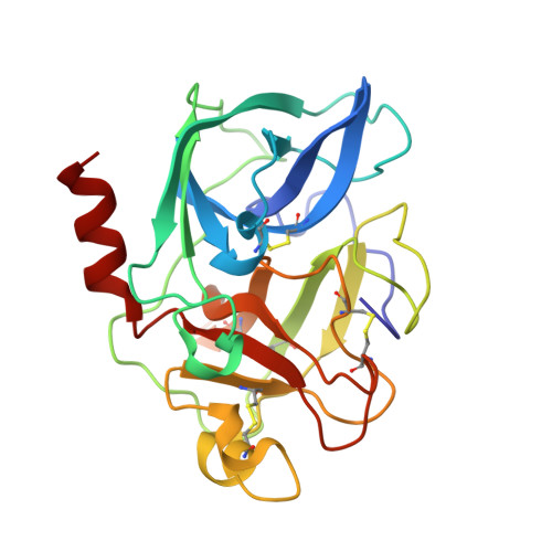
    beta-Lactams inhibit a range of enzymes via acylation of nucleophilic serine residues. Certain gamma-lactam analogues of monocyclic beta-lactams have also been shown to be reversible inhibitors of porcine pancreatic elastase (PPE), forming acyl-enzyme complexes that are stable with respect to hydrolysis. Crystallographic analysis at pH 5 of an acyl-enzyme complex formed with PPE and one of these inhibitors revealed the ester carbonyl located in the oxyanion hole in a similar conformation to that observed in the structure of a complex formed between a heptapeptide (beta-casomorphin-7) and PPE. Only weak electron density was observed for the His-57 side chain in its 'native' conformation. Instead, the His-57 side chain predominantly adopted a conformation rotated approx. 90 degrees from its normal position. PPE-gamma-lactam crystals were subjected to 'pH-jumps' by placing the crystals in a buffer of increased pH prior to freezing for data collection. The results indicate that the conformation of the gamma-lactam-derived acyl-enzyme species in the PPE active site is dependent on pH, a result having implications for the analysis of other serine protease-inhibitor structures at non-catalytic pH values. The results help to define the stereoelectronic relationship between the ester of the acyl-enzyme complex, the side chain of His-57 and the incoming nucleophile during the reversible (de)acylation steps, implying it is closely analogous to the hydrolytic deacylation step during catalytic peptide hydrolysis.


  • Organizational Affiliation
    • The Oxford Centre for Molecular Sciences and The Dyson Perrins Laboratory, South Parks Road, Oxford OX1 3QY, UK.

Macromolecule Content 

  • Total Structure Weight: 26.41 kDa 
  • Atom Count: 2,007 
  • Modeled Residue Count: 240 
  • Deposited Residue Count: 240 
  • Unique protein chains: 1

Macromolecules

Find similar proteins by:|  3D Structure
Entity ID: 1
MoleculeChains  Sequence LengthOrganismDetailsImage
ELASTASEA [auth B]240Sus scrofaMutation(s): 0 
EC: 3.4.21.36
UniProt
Find proteins for P00772 (Sus scrofa)
Explore P00772 
Go to UniProtKB:  P00772
Entity Groups
Sequence Clusters30% Identity50% Identity70% Identity90% Identity95% Identity100% Identity
UniProt GroupP00772
Sequence Annotations
Expand
Reference Sequence

Small Molecules

Ligands 3 Unique
IDChains Name / Formula / InChI Key2D Diagram3D Interactions
TPY

Query on TPY


Download:Ideal Coordinates CCD File
D [auth B](2S,3S)-3-FORMYL-2-({[(4-NITROPHENYL)SULFONYL]AMINO}METHYL)PENTANOIC ACID
C13 H16 N2 O7 S
JPQYVEFTAZEPOD-BXKDBHETSA-N
SO4

Query on SO4


Download:Ideal Coordinates CCD File
C [auth B]SULFATE ION
O4 S
QAOWNCQODCNURD-UHFFFAOYSA-L
CA

Query on CA


Download:Ideal Coordinates CCD File
B
CALCIUM ION
Ca
BHPQYMZQTOCNFJ-UHFFFAOYSA-N

Experimental Data & Validation

Experimental Data

  • Method: X-RAY DIFFRACTION
  • Resolution: 1.70 Å
  • R-Value Free:  0.220 (Depositor) 
  • R-Value Work:  0.183 (Depositor) 
Space Group: P 21 21 21
Unit Cell:
Length ( Å )Angle ( ˚ )
a = 51.07α = 90
b = 57.91β = 90
c = 75.59γ = 90
Software Package:
Software NamePurpose
REFMACrefinement
MOSFLMdata reduction
SCALAdata scaling
X-PLORphasing

Structure Validation

View Full Validation Report


Entry History 

Deposition Data

Revision History  (Full details and data files)

  • Version 1.0: 2000-10-18
    Type: Initial release
  • Version 1.1: 2011-05-08
    Changes: Version format compliance
  • Version 1.2: 2011-07-13
    Changes: Version format compliance
  • Version 1.3: 2023-12-06
    Changes: Data collection, Database references, Derived calculations, Refinement description
  • Version 1.4: 2024-11-20
    Changes: Structure summary