1E0J | pdb_00001e0j

gp4d helicase from phage T7 ADPNP complex


Experimental Data Snapshot

  • Method: X-RAY DIFFRACTION
  • Resolution: 3.00 Å
  • R-Value Free: 
    0.304 (Depositor) 
  • R-Value Work: 
    0.253 (Depositor), 0.240 (DCC) 
  • R-Value Observed: 
    0.253 (Depositor) 

wwPDB Validation   3D Report Full Report


Ligand Structure Quality Assessment 

Created with Raphaël 2.3.0Worse 01 BetterLigand structure goodness of fit to experimental dataBest fitted ANPClick on this verticalbar to view details

This is version 1.3 of the entry. See complete history


Literature

Crystal Structure of T7 Gene 4 Ring Helicase Indicates a Mechanism for Sequential Hydrolysis of Nucleotides

Singleton, M.R.Sawaya, M.R.Ellenberger, T.Wigley, D.B.

(2000) Cell 101: 589

  • DOI: https://doi.org/10.1016/s0092-8674(00)80871-5
  • Primary Citation of Related Structures:  
    1E0J, 1E0K

  • PubMed Abstract: 

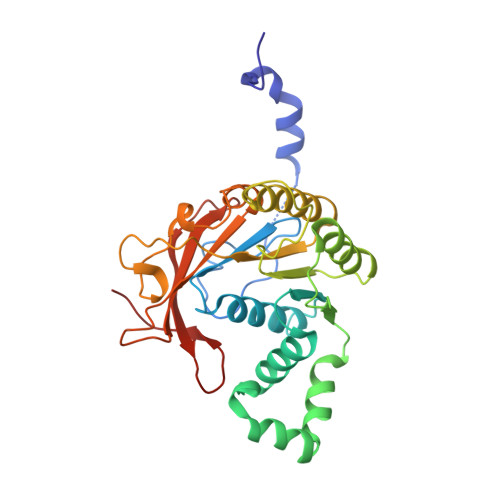
    We have determined the crystal structure of an active, hexameric fragment of the gene 4 helicase from bacteriophage T7. The structure reveals how subunit contacts stabilize the hexamer. Deviation from expected six-fold symmetry of the hexamer indicates that the structure is of an intermediate on the catalytic pathway. The structural consequences of the asymmetry suggest a "binding change" mechanism to explain how cooperative binding and hydrolysis of nucleotides are coupled to conformational changes in the ring that most likely accompany duplex unwinding. The structure of a complex with a nonhydrolyzable ATP analog provides additional evidence for this hypothesis, with only four of the six possible nucleotide binding sites being occupied in this conformation of the hexamer. This model suggests a mechanism for DNA translocation.


  • Organizational Affiliation

    Sir William Dunn School of Pathology, University of Oxford, United Kingdom.


Macromolecules
Find similar proteins by:  (by identity cutoff)  |  3D Structure
Entity ID: 1
MoleculeChains Sequence LengthOrganismDetailsImage
DNA HELICASE
A, B, C, D, E
289Escherichia phage T7Mutation(s): 0 
Gene Names: GENE 4
EC: 3.6.4.12 (UniProt), 2.7.7 (UniProt)
UniProt
Find proteins for P03692 (Escherichia phage T7)
Explore P03692 
Go to UniProtKB:  P03692
Entity Groups  
Sequence Clusters30% Identity50% Identity70% Identity90% Identity95% Identity100% Identity
UniProt GroupP03692
Sequence Annotations
Expand
  • Reference Sequence
Small Molecules
Ligands 2 Unique
IDChains Name / Formula / InChI Key2D Diagram3D Interactions
ANP
Query on ANP

Download Ideal Coordinates CCD File 
G [auth A],
I [auth B],
K [auth D],
M [auth E]
PHOSPHOAMINOPHOSPHONIC ACID-ADENYLATE ESTER
C10 H17 N6 O12 P3
PVKSNHVPLWYQGJ-KQYNXXCUSA-N
MG
Query on MG

Download Ideal Coordinates CCD File 
H [auth A],
J [auth B],
L [auth D],
N [auth E]
MAGNESIUM ION
Mg
JLVVSXFLKOJNIY-UHFFFAOYSA-N
Experimental Data & Validation

Experimental Data

  • Method: X-RAY DIFFRACTION
  • Resolution: 3.00 Å
  • R-Value Free:  0.304 (Depositor) 
  • R-Value Work:  0.253 (Depositor), 0.240 (DCC) 
  • R-Value Observed: 0.253 (Depositor) 
Space Group: P 41 21 2
Unit Cell:
Length ( Å )Angle ( ˚ )
a = 119.18α = 90
b = 119.18β = 90
c = 283.42γ = 90
Software Package:
Software NamePurpose
CNSrefinement
DENZOdata reduction
SCALEPACKdata scaling
CNSphasing

Structure Validation

View Full Validation Report


Ligand Structure Quality Assessment 

Created with Raphaël 2.3.0Worse 01 BetterLigand structure goodness of fit to experimental dataBest fitted ANPClick on this verticalbar to view details

Entry History 

Deposition Data

Revision History  (Full details and data files)

  • Version 1.0: 2000-06-09
    Type: Initial release
  • Version 1.1: 2011-05-08
    Changes: Version format compliance
  • Version 1.2: 2011-07-13
    Changes: Version format compliance
  • Version 1.3: 2024-05-08
    Changes: Data collection, Database references, Derived calculations, Other