1DWU | pdb_00001dwu

Ribosomal protein L1


Experimental Data Snapshot

  • Method: X-RAY DIFFRACTION
  • Resolution: 2.80 Å
  • R-Value Free: 
    0.305 (Depositor) 
  • R-Value Work: 
    0.207 (Depositor), 0.209 (DCC) 
  • R-Value Observed: 
    0.207 (Depositor) 

Starting Model: experimental
View more details

wwPDB Validation 3D Report Full Report

Validation slider image for 1DWU

This is version 1.4 of the entry. See complete history

Literature

Structure of Ribosomal Protein L1 from Methanococcus Thermolithotrophicus. Functionally Important Structural Invariants on the L1 Surface

Nevskaya, N.A.Tishchenko, S.V.Paveliev, M.Smolinskaya, Y.Fedorov, R.Piendl, W.Nakamura, Y.Toyoda, T.Garber, M.B.Nikonov, S.V.

(2002) Acta Crystallogr D Biol Crystallogr 58: 1023

  • DOI: https://doi.org/10.1107/s0907444902006157
  • Primary Citation Related Structures: 
    1DWU

  • PubMed Abstract: 

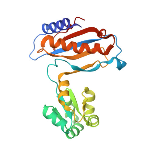
    The crystal structure of ribosomal protein L1 from the archaeon Methanococcus thermolithotrophicus has been determined at 2.7 A resolution. The crystals belong to space group P2(1)2(1)2(1), with unit-cell parameters a = 67.0, b = 70.1, c = 106.3 A and two molecules per asymmetric unit. The structure was solved by the molecular-replacement method with AMoRe and refined with CNS to an R value of 18.9% and an R(free) of 25.4% in the resolution range 30-2.7 A. Comparison of this structure with those obtained previously for two L1 proteins from other sources (the bacterium Thermus thermophilus and the archaeon M. jannaschii) as well as detailed analysis of intermolecular contacts in the corresponding L1 crystals reveal structural invariants on the molecular surface which are probably important for binding the 23S ribosomal RNA and protein function within the ribosome.


  • Organizational Affiliation
    • Institute of Protein Research, Russian Academy of Sciences, 142290 Pushchino, Moscow Region, Russia.

Macromolecule Content 

  • Total Structure Weight: 47.66 kDa 
  • Atom Count: 3,336 
  • Modeled Residue Count: 426 
  • Deposited Residue Count: 426 
  • Unique protein chains: 1

Macromolecules

Find similar proteins by:|  3D Structure
Entity ID: 1
MoleculeChains  Sequence LengthOrganismDetailsImage
RIBOSOMAL PROTEIN L1
A, B
213Methanothermococcus thermolithotrophicusMutation(s): 0 
Gene Names: THE CORRESPONDING GENE IN E. COLI IS RPLA
UniProt
Find proteins for O52704 (Methanothermococcus thermolithotrophicus)
Explore O52704 
Go to UniProtKB:  O52704
Entity Groups
Sequence Clusters30% Identity50% Identity70% Identity90% Identity95% Identity100% Identity
UniProt GroupO52704
Sequence Annotations
Expand
Reference Sequence

Experimental Data & Validation

Experimental Data

  • Method: X-RAY DIFFRACTION
  • Resolution: 2.80 Å
  • R-Value Free:  0.305 (Depositor) 
  • R-Value Work:  0.207 (Depositor), 0.209 (DCC) 
  • R-Value Observed: 0.207 (Depositor) 
Space Group: P 21 21 21
Unit Cell:
Length ( Å )Angle ( ˚ )
a = 67.36α = 90
b = 69.98β = 90
c = 106.17γ = 90
Software Package:
Software NamePurpose
X-PLORrefinement
DENZOdata reduction
SCALEPACKdata scaling
X-PLORphasing

Structure Validation

View Full Validation Report


Entry History 

Deposition Data

Revision History  (Full details and data files)

  • Version 1.0: 2000-12-07
    Type: Initial release
  • Version 1.1: 2011-05-07
    Changes: Version format compliance
  • Version 1.2: 2011-07-13
    Changes: Version format compliance
  • Version 1.3: 2017-07-05
    Changes: Data collection
  • Version 1.4: 2023-12-06
    Changes: Data collection, Database references, Other, Refinement description