1DWE

Crystallographic analysis at 3.0-Angstroms resolution of the binding to human thrombin of four active site-directed inhibitors


Experimental Data Snapshot

  • Method: X-RAY DIFFRACTION
  • Resolution: 3.00 Å
  • R-Value Observed: 0.164 

wwPDB Validation   3D Report Full Report


This is version 1.5 of the entry. See complete history


Literature

Crystallographic analysis at 3.0-A resolution of the binding to human thrombin of four active site-directed inhibitors.

Banner, D.W.Hadvary, P.

(1991) J Biol Chem 266: 20085-20093

  • Primary Citation of Related Structures:  
    1DWB, 1DWC, 1DWD, 1DWE

  • PubMed Abstract: 

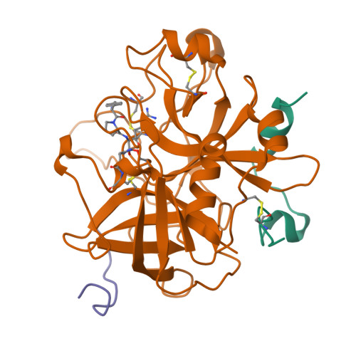
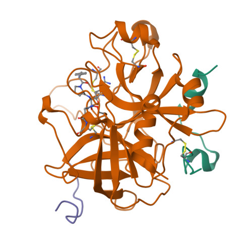
    The mode of binding of four active-site directed inhibitors to human thrombin has been determined by x-ray crystallographic analysis. The inhibitors studied are benzamidine, PPACK, NAPAP, and MD-805, of which the last three are compounds evolved specifically to inhibit thrombin. Crystal structures were determined in the presence of both the inhibitor and the undecapeptide [des-amino Asp55]hirudin(55-65) which binds distant from the active site. Despite having significantly different chemical structures, NAPAP and MD-805 bind to thrombin in a very similar "inhibitor binding mode" which is not that expected by direct analogy with the binding of substrate. Both inhibitors bind to thrombin in a similar way as to trypsin, but thrombin has an extra loop, the "Tyr-Pro-Pro-Trp loop," not present in trypsin, which gives further binding interactions and is seen to move somewhat to accommodate binding of the different inhibitors. The fact that NAPAP and MD-805 require different stereochemistry for potent inhibition is demonstrated, and its structural basis clarified. The wealth of data on analogs and variants of these lead compounds is shown to be compatible with this inhibitor binding mode.


  • Organizational Affiliation

    Pharmaceutical Research Departments, F. Hoffmann-La Roche Ltd., Basel, Switzerland.


Macromolecules
Find similar proteins by:  (by identity cutoff)  |  3D Structure
Entity ID: 1
MoleculeChains Sequence LengthOrganismDetailsImage
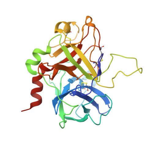
ALPHA-THROMBIN light chainA [auth L]36Homo sapiensMutation(s): 0 
EC: 3.4.21.5
UniProt & NIH Common Fund Data Resources
Find proteins for P00734 (Homo sapiens)
Explore P00734 
Go to UniProtKB:  P00734
PHAROS:  P00734
GTEx:  ENSG00000180210 
Entity Groups  
Sequence Clusters30% Identity50% Identity70% Identity90% Identity95% Identity100% Identity
UniProt GroupP00734
Sequence Annotations
Expand
  • Reference Sequence
Find similar proteins by:  (by identity cutoff)  |  3D Structure
Entity ID: 2
MoleculeChains Sequence LengthOrganismDetailsImage
ALPHA-THROMBIN heavy chainB [auth H]259Homo sapiensMutation(s): 0 
EC: 3.4.21.5
UniProt & NIH Common Fund Data Resources
Find proteins for P00734 (Homo sapiens)
Explore P00734 
Go to UniProtKB:  P00734
PHAROS:  P00734
GTEx:  ENSG00000180210 
Entity Groups  
Sequence Clusters30% Identity50% Identity70% Identity90% Identity95% Identity100% Identity
UniProt GroupP00734
Sequence Annotations
Expand
  • Reference Sequence

Find similar proteins by:  Sequence   |   3D Structure  

Entity ID: 3
MoleculeChains Sequence LengthOrganismDetailsImage
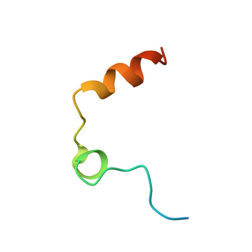
HIRUDIN IIIAC [auth I]11Hirudo medicinalisMutation(s): 0 
UniProt
Find proteins for P28508 (Hirudo medicinalis)
Explore P28508 
Go to UniProtKB:  P28508
Entity Groups  
Sequence Clusters30% Identity50% Identity70% Identity90% Identity95% Identity100% Identity
UniProt GroupP28508
Sequence Annotations
Expand
  • Reference Sequence
Small Molecules
Ligands 1 Unique
IDChains Name / Formula / InChI Key2D Diagram3D Interactions
0G6
Query on 0G6

Download Ideal Coordinates CCD File 
D [auth H]D-phenylalanyl-N-[(2S,3S)-6-{[amino(iminio)methyl]amino}-1-chloro-2-hydroxyhexan-3-yl]-L-prolinamide
C21 H34 Cl N6 O3
DVFLYEYCMMLBTQ-VSZNYVQBSA-O
Biologically Interesting Molecules (External Reference) 1 Unique
Entity ID: 4
IDChains NameType/Class2D Diagram3D Interactions
PRD_000020 (0G6)
Query on PRD_000020
D [auth H]D-Phe-Pro-Arg-CH2ClPeptide-like / Inhibitor
Experimental Data & Validation

Experimental Data

  • Method: X-RAY DIFFRACTION
  • Resolution: 3.00 Å
  • R-Value Observed: 0.164 
  • Space Group: P 43 21 2
Unit Cell:
Length ( Å )Angle ( ˚ )
a = 90.8α = 90
b = 90.8β = 90
c = 132.5γ = 90
Software Package:
Software NamePurpose
TNTrefinement

Structure Validation

View Full Validation Report


Entry History 

Deposition Data

Revision History  (Full details and data files)

  • Version 1.0: 1994-01-31
    Type: Initial release
  • Version 1.1: 2008-03-06
    Changes: Version format compliance
  • Version 1.2: 2011-07-13
    Changes: Atomic model, Database references, Derived calculations, Non-polymer description, Structure summary, Version format compliance
  • Version 1.3: 2013-02-27
    Changes: Other
  • Version 1.4: 2024-06-05
    Changes: Data collection, Database references, Derived calculations, Other
  • Version 1.5: 2024-10-16
    Changes: Structure summary