1DUD | pdb_00001dud

DEOXYURIDINE 5'-TRIPHOSPHATE NUCLEOTIDE HYDROLASE (D-UTPASE) COMPLEXED WITH THE SUBSTRATE ANALOGUE DEOXYURIDINE 5'-DIPHOSPHATE (D-UDP)


Experimental Data Snapshot

  • Method: X-RAY DIFFRACTION
  • Resolution: 2.30 Å
  • R-Value Work: 
    0.200 (Depositor), 0.174 (DCC) 
  • R-Value Observed: 
    0.200 (Depositor) 

wwPDB Validation 3D Report Full Report

Validation slider image for 1DUD

Ligand Structure Quality Assessment 


This is version 1.4 of the entry. See complete history

Literature

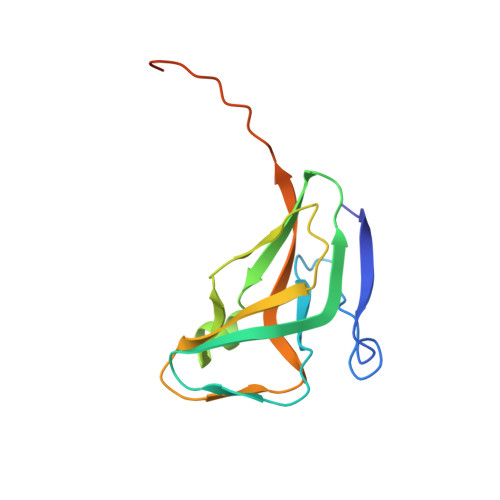
Crystal structure of the Escherichia coli dUTPase in complex with a substrate analogue (dUDP).

Larsson, G.Svensson, L.A.Nyman, P.O.

(1996) Nat Struct Biol 3: 532-538

  • DOI: https://doi.org/10.1038/nsb0696-532
  • Primary Citation Related Structures: 
    1DUD

  • PubMed Abstract: 

    We have determined the structure of the homotrimeric dUTPase from Escherichia coli, completed with an inhibitor and substrate analogue, dUDP. Three molecules of dUDP are found symmetrically bound per trimer, each in a shallow cleft between adjacent subunits, interacting with evolutionary conserved residues. The interactions of the uracil ring and the deoxypentose with the protein are consistent with the high specificity of the enzyme with respect to these groups. The positions of the two phosphate groups and adjacent water molecules are discussed in relation to the mechanism and kinetics of catalysis. The role that dUTPase plays in DNA metabolism makes the enzyme a potential target for chemotherapeutic drugs: the results presented here will aid in the design and development of inhibitory compounds.


  • Organizational Affiliation
    • Department of Biochemistry, Center for Chemistry and Chemical Engineering, Lund University, Sweden.

Macromolecule Content 

  • Total Structure Weight: 16.69 kDa 
  • Atom Count: 1,145 
  • Modeled Residue Count: 136 
  • Deposited Residue Count: 152 
  • Unique protein chains: 1

Macromolecules

Find similar proteins by:|  3D Structure
Entity ID: 1
MoleculeChains  Sequence LengthOrganismDetailsImage
DEOXYURIDINE 5'-TRIPHOSPHATE NUCLEOTIDOHYDROLASE152Escherichia coliMutation(s): 0 
EC: 3.6.1.23
UniProt
Find proteins for P06968 (Escherichia coli (strain K12))
Explore P06968 
Go to UniProtKB:  P06968
Entity Groups
Sequence Clusters30% Identity50% Identity70% Identity90% Identity95% Identity100% Identity
UniProt GroupP06968
Sequence Annotations
Expand
Reference Sequence

Small Molecules

Ligands 1 Unique
IDChains Name / Formula / InChI Key2D Diagram3D Interactions
DUD

Query on DUD


Download:Ideal Coordinates CCD File
B [auth A]DEOXYURIDINE-5'-DIPHOSPHATE
C9 H14 N2 O11 P2
QHWZTVCCBMIIKE-SHYZEUOFSA-N

Experimental Data & Validation

Experimental Data

  • Method: X-RAY DIFFRACTION
  • Resolution: 2.30 Å
  • R-Value Work:  0.200 (Depositor), 0.174 (DCC) 
  • R-Value Observed: 0.200 (Depositor) 
Space Group: H 3
Unit Cell:
Length ( Å )Angle ( ˚ )
a = 86.64α = 90
b = 86.64β = 90
c = 62.23γ = 120
Software Package:
Software NamePurpose
X-PLORmodel building
X-PLORrefinement
XDSdata reduction
X-PLORphasing

Structure Validation

View Full Validation Report


Ligand Structure Quality Assessment 


Entry History 

Deposition Data

Revision History  (Full details and data files)

  • Version 1.0: 1996-11-08
    Type: Initial release
  • Version 1.1: 2008-03-24
    Changes: Version format compliance
  • Version 1.2: 2011-07-13
    Changes: Derived calculations, Version format compliance
  • Version 1.3: 2017-11-29
    Changes: Derived calculations, Other
  • Version 1.4: 2024-02-07
    Changes: Data collection, Database references, Derived calculations