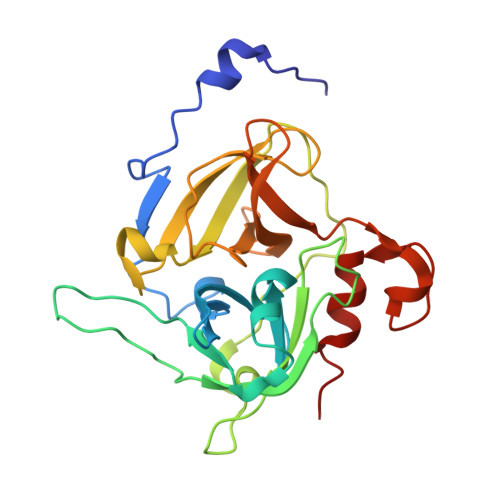
Structural similarities and differences in Staphylococcus aureus exfoliative toxins A and B as revealed by their crystal structures.
Papageorgiou, A.C., Plano, L.R., Collins, C.M., Acharya, K.R.(2000) Protein Sci 9: 610-618
- PubMed: 10752623
- DOI: https://doi.org/10.1110/ps.9.3.610
- Primary Citation of Related Structures:
1DT2, 1DUA, 1DUE - PubMed Abstract:
Staphylococcal aureus epidermolytic toxins (ETs) A and B are responsible for the induction of staphylococcal scalded skin syndrome, a disease of neonates and young children. The clinical features of this syndrome vary from localized blisters to severe exfoliation affecting most of the body surface. Comparison of the crystal structures of two subtypes of ETs-rETA (at 2.0 A resolution), rETB (at 2.8 A resolution), and an active site variant of rETA, Ser195Ala at 2.0 A resolution has demonstrated that their overall topology resembles that of a "trypsin-like" serine protease, but with significant differences at the N- and C-termini and loop regions. The details of the catalytic site in both ET structures are very similar to those in glutamate-specific serine proteases, suggesting a common catalytic mechanism. However, the "oxyanion hole," which is part of the catalytic sites of glutamate specific serine proteases, is in the closed or inactive conformation for rETA, yet in the open or active conformation for rETB. The ETs contain a unique amphipathic helix at the N-terminus, and it appears to be involved in optimizing the conformation of the catalytic site residues. Determination of the structure of the rETA catalytic site variant, Ser195Ala, showed no significant perturbation at the active site, establishing that the loss of biological and esterolytic activity can be attributed solely to disruption of the catalytic serine residue. Finally, the crystal structure of ETs, together with biochemical data and mutagenesis studies, strongly confirms the classification of these molecules as "serine proteases" rather than "superantigens."
Organizational Affiliation:
Department of Biology and Biochemistry, University of Bath, United Kingdom.


