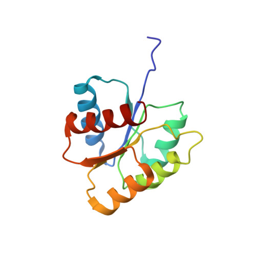
NMR structure of activated CheY.
Cho, H.S., Lee, S.Y., Yan, D., Pan, X., Parkinson, J.S., Kustu, S., Wemmer, D.E., Pelton, J.G.(2000) J Mol Biology 297: 543-551
- PubMed: 10731410 Search on PubMed
- DOI: https://doi.org/10.1006/jmbi.2000.3595
- Primary Citation Related Structures:
1DJM - PubMed Abstract:
The CheY protein is the response regulator in bacterial chemotaxis. Phosphorylation of a conserved aspartyl residue induces structural changes that convert the protein from an inactive to an active state. The short half-life of the aspartyl-phosphate has precluded detailed structural analysis of the active protein. Persistent activation of Escherichia coli CheY was achieved by complexation with beryllofluoride (BeF(3)(-)) and the structure determined by NMR spectroscopy to a backbone r.m.s.d. of 0.58(+/-0.08) A. Formation of a hydrogen bond between the Thr87 OH group and an active site acceptor, presumably Asp57.BeF(3)(-), stabilizes a coupled rearrangement of highly conserved residues, Thr87 and Tyr106, along with displacement of beta4 and H4, to yield the active state. The coupled rearrangement may be a more general mechanism for activation of receiver domains.
- Physical Biosciences Division, Lawrence Berkeley National Laboratory, 1 Cyclotron Rd, Berkeley, CA, 94720, USA.
Organizational Affiliation: