1DJ0 | pdb_00001dj0

THE CRYSTAL STRUCTURE OF E. COLI PSEUDOURIDINE SYNTHASE I AT 1.5 ANGSTROM RESOLUTION


Experimental Data Snapshot

  • Method: X-RAY DIFFRACTION
  • Resolution: 1.50 Å
  • R-Value Free: 
    0.221 (Depositor) 
  • R-Value Work: 
    0.167 (Depositor) 
  • R-Value Observed: 
    0.162 (Depositor) 

wwPDB Validation 3D Report Full Report

Validation slider image for 1DJ0

This is version 1.3 of the entry. See complete history

Literature

The structural basis for tRNA recognition and pseudouridine formation by pseudouridine synthase I.

Foster, P.G.Huang, L.Santi, D.V.Stroud, R.M.

(2000) Nat Struct Biol 7: 23-27

  • DOI: https://doi.org/10.1038/71219
  • Primary Citation Related Structures: 
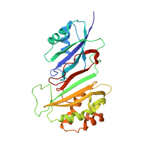
    1DJ0

  • PubMed Abstract: 

    Pseudouridine synthases catalyze the isomerization of specific uridines to pseudouridine in a variety of RNAs, yet the basis for recognition of the RNA sites or how they catalyze this reaction is unknown. The crystal structure of pseudouridine synthase I from Escherichia coli, which, for example, modifies positions 38, 39 and/or 40 in tRNA, reveals a dimeric protein that contains two positively charged, RNA-binding clefts along the surface of the protein. Each cleft contains a highly conserved aspartic acid located at its center. The structural domains have a topological similarity to those of other RNA-binding proteins, though the mode of interaction with tRNA appears to be unique. The structure suggests that a dimeric enzyme is required for binding transfer RNA and subsequent pseudouridine formation.


  • Organizational Affiliation
    • Department of Biochemistry and Biophysics, University of California San Francisco, San Francisco, California 94143-0448, USA.

Macromolecule Content 

  • Total Structure Weight: 59.52 kDa 
  • Atom Count: 4,663 
  • Modeled Residue Count: 528 
  • Deposited Residue Count: 528 
  • Unique protein chains: 1

Macromolecules

Find similar proteins by:|  3D Structure
Entity ID: 1
MoleculeChains  Sequence LengthOrganismDetailsImage
PSEUDOURIDINE SYNTHASE I
A, B
264Escherichia coliMutation(s): 0 
EC: 4.2.1.70 (PDB Primary Data), 5.4.99.12 (UniProt)
UniProt
Find proteins for P07649 (Escherichia coli (strain K12))
Explore P07649 
Go to UniProtKB:  P07649
Entity Groups
Sequence Clusters30% Identity50% Identity70% Identity90% Identity95% Identity100% Identity
UniProt GroupP07649
Sequence Annotations
Expand
Reference Sequence

Small Molecules

Experimental Data & Validation

Experimental Data

  • Method: X-RAY DIFFRACTION
  • Resolution: 1.50 Å
  • R-Value Free:  0.221 (Depositor) 
  • R-Value Work:  0.167 (Depositor) 
  • R-Value Observed: 0.162 (Depositor) 
Space Group: P 1 21 1
Unit Cell:
Length ( Å )Angle ( ˚ )
a = 68.08α = 90
b = 37.4β = 97.68
c = 103.48γ = 90
Software Package:
Software NamePurpose
PHASESphasing
REFMACrefinement
DENZOdata reduction
SCALEPACKdata scaling

Structure Validation

View Full Validation Report


Entry History 

Deposition Data

Revision History  (Full details and data files)

  • Version 1.0: 2000-05-31
    Type: Initial release
  • Version 1.1: 2008-04-27
    Changes: Version format compliance
  • Version 1.2: 2011-07-13
    Changes: Version format compliance
  • Version 1.3: 2024-02-07
    Changes: Data collection, Database references, Derived calculations