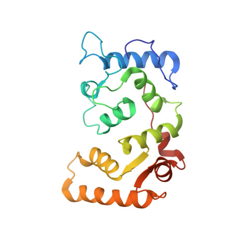
Structures of the platelet calcium- and integrin-binding protein and the alphaIIb-integrin cytoplasmic domain suggest a mechanism for calcium-regulated recognition; homology modelling and NMR studies.
Hwang, P.M., Vogel, H.J.(2000) J Mol Recognit 13: 83-92
- PubMed: 10822252
- DOI: https://doi.org/10.1002/(SICI)1099-1352(200003/04)13:2<83::AID-JMR491>3.0.CO;2-A
- Primary Citation of Related Structures:
1DGU, 1DGV - PubMed Abstract:
Calcium- and integrin-binding protein (CIB) binds to the 20-residue alphaIIb cytoplasmic domain of platelet alphaIIbbeta3 integrin. Amino acid sequence similarities with calmodulin (CaM) and calcineurin B (CnB) allowed the construction of homology-based models of calcium-saturated CIB as well as apo-CIB. In addition, the solution structure of the alphaIIb cytoplasmic domain in 45% aqueous trifluoroethanol was solved by conventional two-dimensional NMR methods. The models indicate that the N-terminal domain of CIB possesses a number of positively charged residues in its binding site that could interact with the acidic carboxy-terminal LEEDDEEGE sequence of alphaIIb. The C-terminal domain of CIB seems well-suited to bind the sequence WKVGFFKR, which forms a well-structured alpha helix; this is analogous to calmodulin and calcineurin B, which also bind alpha helices. Similarities between the C-terminal domains of CIB and calmodulin suggest that binding of CIB to the cytoplasmic domain of alphaIIb may be affected by fluctuations in the intracellular calcium concentration.
Organizational Affiliation:
Department of Biological Sciences, University of Calgary, Alberta, Canada.


