1DG1 | pdb_00001dg1

WHOLE, UNMODIFIED, EF-TU(ELONGATION FACTOR TU).


Experimental Data Snapshot

  • Method: X-RAY DIFFRACTION
  • Resolution: 2.50 Å
  • R-Value Free: 
    0.259 (Depositor) 
  • R-Value Work: 
    0.168 (Depositor) 
  • R-Value Observed: 
    0.168 (Depositor) 

wwPDB Validation 3D Report Full Report

Validation slider image for 1DG1

This is version 1.3 of the entry. See complete history

Literature

An alpha to beta conformational switch in EF-Tu.

Abel, K.Yoder, M.D.Hilgenfeld, R.Jurnak, F.

(1996) Structure 4: 1153-1159

  • DOI: https://doi.org/10.1016/s0969-2126(96)00123-2
  • Primary Citation Related Structures: 
    1DG1

  • PubMed Abstract: 

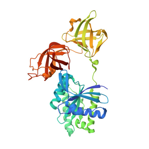
    The bacterial elongation factor EF-Tu recognizes and transports aminoacyl-tRNAs to mRNA-programmed ribosomes. EF-Tu shares many structural and functional properties with other GTPases whose conformations are regulated by guanine nucleotides. An intact form of Escherichia coli EF-Tu complexed with GDP has been crystallized in the presence of the EF-Tu-specific antibiotic GE2270 A. The three-dimensional structure has been solved by X-ray diffraction analysis and refined to a final crystallographic R factor of 17.2% at a resolution of 2.5 A. The location of the GE2270 A antibiotic-binding site could not be identified. The structure of EF-Tu-GDP is nearly identical to that of a trypsin-modified form of EF-Tu-GDP, demonstrating conclusively that the protease treatment had not altered any essential structural features. The present structure represents the first view of an ordered Switch I region in EF-Tu-GDP and reveals similarities with two other GTPases complexed with GDP: Ran and ADP-ribosylation factor-1. A comparison of the Switch I regions of the GTP and GDP forms of EF-Tu also reveals that a segment, six amino acids in length, completely converts from an alpha helix in the GTP complex to beta secondary structure in the GDP form. The alpha to beta switch in EF-Tu may represent a prototypical activation mechanism for other protein families.


  • Organizational Affiliation
    • Department of Biochemistry, University of California, Riverside, CA 92521-0129, USA.

Macromolecule Content 

  • Total Structure Weight: 87.68 kDa 
  • Atom Count: 6,177 
  • Modeled Residue Count: 770 
  • Deposited Residue Count: 788 
  • Unique protein chains: 1

Macromolecules

Find similar proteins by:|  3D Structure
Entity ID: 1
MoleculeChains  Sequence LengthOrganismDetailsImage
ELONGATION FACTOR TUA [auth G],
B [auth H]
394Escherichia coliMutation(s): 0 
EC: 3.6.5.3
UniProt
Find proteins for P0CE48 (Escherichia coli (strain K12))
Explore P0CE48 
Go to UniProtKB:  P0CE48
Entity Groups
Sequence Clusters30% Identity50% Identity70% Identity90% Identity95% Identity100% Identity
UniProt GroupP0CE48
Sequence Annotations
Expand
Reference Sequence

Experimental Data & Validation

Experimental Data

  • Method: X-RAY DIFFRACTION
  • Resolution: 2.50 Å
  • R-Value Free:  0.259 (Depositor) 
  • R-Value Work:  0.168 (Depositor) 
  • R-Value Observed: 0.168 (Depositor) 
Space Group: P 1 21 1
Unit Cell:
Length ( Å )Angle ( ˚ )
a = 63.6α = 90
b = 104.6β = 97.02
c = 67.2γ = 90
Software Package:
Software NamePurpose
X-PLORmodel building
X-PLORrefinement
SDMSdata reduction
SDMSdata scaling
X-PLORphasing

Structure Validation

View Full Validation Report


Entry History 

Deposition Data

Revision History  (Full details and data files)

  • Version 1.0: 1999-12-01
    Type: Initial release
  • Version 1.1: 2008-04-27
    Changes: Version format compliance
  • Version 1.2: 2011-07-13
    Changes: Version format compliance
  • Version 1.3: 2024-02-07
    Changes: Data collection, Database references, Derived calculations