1DCC | pdb_00001dcc

2.2 ANGSTROM STRUCTURE OF OXYPEROXIDASE: A MODEL FOR THE ENZYME:PEROXIDE COMPLEX


Experimental Data Snapshot

  • Method: X-RAY DIFFRACTION
  • Resolution: 2.20 Å
  • R-Value Observed: 
    0.155 (Depositor) 

wwPDB Validation 3D Report Full Report

Validation slider image for 1DCC

This is version 1.4 of the entry. See complete history

Literature

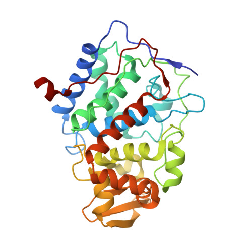
2.2 A structure of oxy-peroxidase as a model for the transient enzyme: peroxide complex.

Miller, M.A.Shaw, A.Kraut, J.

(1994) Nat Struct Biol 1: 524-531

  • DOI: https://doi.org/10.1038/nsb0894-524
  • Primary Citation Related Structures: 
    1DCC

  • PubMed Abstract: 

    The Fe+3-OOH complex of peroxidases has a very short half life, and its structure cannot be determined by conventional methods. The Fe+2-O2 complex provides a useful structural model for this intermediate, as it differs by only one electron and one proton from the transient Fe+3-OOH complex. We therefore determined the crystal structure of the Fe+2-O2 complex formed by a yeast cytochrome c peroxidase mutant with Trp 191 replaced by Phe. The refined structure shows that dioxygen can form a hydrogen bond with the conserved distal His residue, but not with the conserved distal Arg residue. When the transient Fe+3-OOH complex is modelled in a similar orientation, the active site of peroxidase appears to be optimized for catalysing proton transfer between the vicinal oxygen atoms of the peroxy-anion.


  • Organizational Affiliation
    • Department of Chemistry, University of California, San Diego La Jolla 92093-0317.

Macromolecule Content 

  • Total Structure Weight: 34.38 kDa 
  • Atom Count: 2,552 
  • Modeled Residue Count: 291 
  • Deposited Residue Count: 296 
  • Unique protein chains: 1

Macromolecules

Find similar proteins by:|  3D Structure
Entity ID: 1
MoleculeChains  Sequence LengthOrganismDetailsImage
CYTOCHROME C PEROXIDASE296Saccharomyces cerevisiaeMutation(s): 0 
EC: 1.11.1.5
UniProt
Find proteins for P00431 (Saccharomyces cerevisiae (strain ATCC 204508 / S288c))
Explore P00431 
Go to UniProtKB:  P00431
Entity Groups
Sequence Clusters30% Identity50% Identity70% Identity90% Identity95% Identity100% Identity
UniProt GroupP00431
Sequence Annotations
Expand
Reference Sequence

Small Molecules

Ligands 2 Unique
IDChains Name / Formula / InChI Key2D Diagram3D Interactions
HEM

Query on HEM


Download:Ideal Coordinates CCD File
C [auth A]PROTOPORPHYRIN IX CONTAINING FE
C34 H32 Fe N4 O4
KABFMIBPWCXCRK-RGGAHWMASA-L
OXY

Query on OXY


Download:Ideal Coordinates CCD File
B [auth A]OXYGEN MOLECULE
O2
MYMOFIZGZYHOMD-UHFFFAOYSA-N

Experimental Data & Validation

Experimental Data

  • Method: X-RAY DIFFRACTION
  • Resolution: 2.20 Å
  • R-Value Observed: 0.155 (Depositor) 
Space Group: P 21 21 21
Unit Cell:
Length ( Å )Angle ( ˚ )
a = 104.722α = 90
b = 74.446β = 90
c = 45.17γ = 90
Software Package:
Software NamePurpose
TNTrefinement

Structure Validation

View Full Validation Report


Entry History 

Deposition Data

Revision History  (Full details and data files)

  • Version 1.0: 1994-08-31
    Type: Initial release
  • Version 1.1: 2008-03-24
    Changes: Version format compliance
  • Version 1.2: 2011-07-13
    Changes: Version format compliance
  • Version 1.3: 2017-11-29
    Changes: Derived calculations, Other
  • Version 1.4: 2024-02-07
    Changes: Data collection, Database references, Derived calculations