1DAK | pdb_00001dak

DETHIOBIOTIN SYNTHETASE FROM ESCHERICHIA COLI, COMPLEX REACTION INTERMEDIATE ADP AND MIXED ANHYDRIDE


Experimental Data Snapshot

  • Method: X-RAY DIFFRACTION
  • Resolution: 1.60 Å
  • R-Value Free: 
    0.210 (Depositor) 
  • R-Value Work: 
    0.184 (Depositor) 

Starting Model: experimental
View more details

wwPDB Validation 3D Report Full Report

Validation slider image for 1DAK

This is version 1.4 of the entry. See complete history

Literature

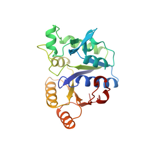
Snapshot of a phosphorylated substrate intermediate by kinetic crystallography.

Kack, H.Gibson, K.J.Lindqvist, Y.Schneider, G.

(1998) Proc Natl Acad Sci U S A 95: 5495-5500

  • DOI: https://doi.org/10.1073/pnas.95.10.5495
  • Primary Citation Related Structures: 
    1A82, 1DAK

  • PubMed Abstract: 

    The ATP-dependent enzyme dethiobiotin synthetase from Escherichia coli catalyses the formation of dethiobiotin from CO2 and 7, 8-diaminopelargonic acid. The reaction is initiated by the formation of a carbamate and proceeds through a phosphorylated intermediate, a mixed carbamic phosphoric anhydride. Here, we report the crystal structures at 1.9- and 1.6-A resolution, respectively, of the enzyme-MgATP-diaminopelargonic acid and enzyme-MgADP-carbamic-phosphoric acid anhydride complexes, observed by using kinetic crystallography. Reaction initiation by addition of either NaHCO3 or diaminopelargonic acid to crystals already containing cosubstrates resulted in the accumulation of the phosphorylated intermediate at the active site. The phosphoryl transfer step shows inversion of the configuration at the phosphorus atom, consistent with an in-line attack by the carbamate oxygen onto the phosphorus atom of ATP. A key feature in the structure of the complex of the enzyme with the reaction intermediate is two magnesium ions, bridging the phosphates at the cleavage site. These magnesium ions compensate the negative charges at both phosphate groups after phosphoryl transfer and contribute to the stabilization of the reaction intermediate.


  • Organizational Affiliation
    • Molecular Structural Biology, Department of Medical Biochemistry and Biophysics, Karolinska Institutet, 171 77 Stockholm, Sweden.

Macromolecule Content 

  • Total Structure Weight: 24.91 kDa 
  • Atom Count: 1,962 
  • Modeled Residue Count: 224 
  • Deposited Residue Count: 224 
  • Unique protein chains: 1

Macromolecules

Find similar proteins by:|  3D Structure
Entity ID: 1
MoleculeChains  Sequence LengthOrganismDetailsImage
DETHIOBIOTIN SYNTHETASE224Escherichia coliMutation(s): 0 
EC: 6.3.3.3
UniProt
Find proteins for P13000 (Escherichia coli (strain K12))
Explore P13000 
Go to UniProtKB:  P13000
Entity Groups
Sequence Clusters30% Identity50% Identity70% Identity90% Identity95% Identity100% Identity
UniProt GroupP13000
Sequence Annotations
Expand
Reference Sequence

Small Molecules

Ligands 4 Unique
IDChains Name / Formula / InChI Key2D Diagram3D Interactions
ADP

Query on ADP


Download:Ideal Coordinates CCD File
F [auth A]ADENOSINE-5'-DIPHOSPHATE
C10 H15 N5 O10 P2
XTWYTFMLZFPYCI-KQYNXXCUSA-N
DPU

Query on DPU


Download:Ideal Coordinates CCD File
E [auth A]MIXED CARBAMIC PHOSPHORIC ACID ANHYDRIDE OF 7,8-DIAMINONONANIC ACID
C10 H21 N2 O7 P
UJJZZEABROBUCE-JGVFFNPUSA-N
PO4

Query on PO4


Download:Ideal Coordinates CCD File
B [auth A]PHOSPHATE ION
O4 P
NBIIXXVUZAFLBC-UHFFFAOYSA-K
MG

Query on MG


Download:Ideal Coordinates CCD File
C [auth A],
D [auth A]
MAGNESIUM ION
Mg
JLVVSXFLKOJNIY-UHFFFAOYSA-N

Experimental Data & Validation

Experimental Data

  • Method: X-RAY DIFFRACTION
  • Resolution: 1.60 Å
  • R-Value Free:  0.210 (Depositor) 
  • R-Value Work:  0.184 (Depositor) 
Space Group: C 1 2 1
Unit Cell:
Length ( Å )Angle ( ˚ )
a = 72.68α = 90
b = 48.19β = 106.58
c = 61.07γ = 90
Software Package:
Software NamePurpose
CCP4model building
REFMACrefinement
DENZOdata reduction
SCALEPACKdata scaling
CCP4phasing

Structure Validation

View Full Validation Report


Entry History 

Deposition Data

Revision History  (Full details and data files)

  • Version 1.0: 1999-04-06
    Type: Initial release
  • Version 1.1: 2008-03-24
    Changes: Version format compliance
  • Version 1.2: 2011-07-13
    Changes: Derived calculations, Version format compliance
  • Version 1.3: 2023-08-09
    Changes: Database references, Derived calculations, Other, Refinement description
  • Version 1.4: 2024-05-22
    Changes: Data collection