1D2T | pdb_00001d2t

CRYSTAL STRUCTURE OF ACID PHOSPHATASE FROM ESCHERICHIA BLATTAE


Experimental Data Snapshot

  • Method: X-RAY DIFFRACTION
  • Resolution: 1.90 Å
  • R-Value Free: 
    0.257 (Depositor) 
  • R-Value Work: 
    0.216 (Depositor), 0.206 (DCC) 
  • R-Value Observed: 
    0.215 (Depositor) 

wwPDB Validation 3D Report Full Report

Validation slider image for 1D2T

This is version 1.3 of the entry. See complete history

Literature

X-ray structures of a novel acid phosphatase from Escherichia blattae and its complex with the transition-state analog molybdate.

Ishikawa, K.Mihara, Y.Gondoh, K.Suzuki, E.Asano, Y.

(2000) EMBO J 19: 2412-2423

  • DOI: https://doi.org/10.1093/emboj/19.11.2412
  • Primary Citation Related Structures: 
    1D2T, 1EOI

  • PubMed Abstract: 

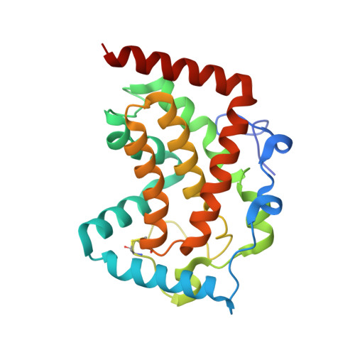
    The structure of Escherichia blattae non-specific acid phosphatase (EB-NSAP) has been determined at 1.9 A resolution with a bound sulfate marking the phosphate-binding site. The enzyme is a 150 kDa homohexamer. EB-NSAP shares a conserved sequence motif not only with several lipid phosphatases and the mammalian glucose-6-phosphatases, but also with the vanadium-containing chloroperoxidase (CPO) of Curvularia inaequalis. Comparison of the crystal structures of EB-NSAP and CPO reveals striking similarity in the active site structures. In addition, the topology of the EB-NSAP core shows considerable similarity to the fold of the active site containing part of the monomeric 67 kDa CPO, despite the lack of further sequence identity. These two enzymes are apparently related by divergent evolution. We have also determined the crystal structure of EB-NSAP complexed with the transition-state analog molybdate. Structural comparison of the native enzyme and the enzyme-molybdate complex reveals that the side-chain of His150, a putative catalytic residue, moves toward the molybdate so that it forms a hydrogen bond with the metal oxyanion when the molybdenum forms a covalent bond with NE2 of His189.


  • Organizational Affiliation
    • Central Research Laboratories, Ajinomoto Co., Inc., 1-1 Suzuki-cho, Kawasaki-ku, Kawasaki 210-8681, Japan.

Macromolecule Content 

  • Total Structure Weight: 25.13 kDa 
  • Atom Count: 1,937 
  • Modeled Residue Count: 222 
  • Deposited Residue Count: 231 
  • Unique protein chains: 1

Macromolecules

Find similar proteins by:|  3D Structure
Entity ID: 1
MoleculeChains  Sequence LengthOrganismDetailsImage
ACID PHOSPHATASE231Shimwellia blattaeMutation(s): 0 
EC: 3.1.3.2
UniProt
Find proteins for Q9S1A6 (Shimwellia blattae)
Explore Q9S1A6 
Go to UniProtKB:  Q9S1A6
Entity Groups
Sequence Clusters30% Identity50% Identity70% Identity90% Identity95% Identity100% Identity
UniProt GroupQ9S1A6
Sequence Annotations
Expand
Reference Sequence

Small Molecules

Ligands 1 Unique
IDChains Name / Formula / InChI Key2D Diagram3D Interactions
SO4

Query on SO4


Download:Ideal Coordinates CCD File
B [auth A]SULFATE ION
O4 S
QAOWNCQODCNURD-UHFFFAOYSA-L

Experimental Data & Validation

Experimental Data

  • Method: X-RAY DIFFRACTION
  • Resolution: 1.90 Å
  • R-Value Free:  0.257 (Depositor) 
  • R-Value Work:  0.216 (Depositor), 0.206 (DCC) 
  • R-Value Observed: 0.215 (Depositor) 
Space Group: P 63 2 2
Unit Cell:
Length ( Å )Angle ( ˚ )
a = 124.7α = 90
b = 124.7β = 90
c = 97.7γ = 120
Software Package:
Software NamePurpose
DENZOdata reduction
SCALAdata scaling
MLPHAREphasing
X-PLORrefinement
CCP4data scaling

Structure Validation

View Full Validation Report


Entry History 

Deposition Data

Revision History  (Full details and data files)

  • Version 1.0: 2000-12-06
    Type: Initial release
  • Version 1.1: 2007-10-16
    Changes: Version format compliance
  • Version 1.2: 2011-07-13
    Changes: Version format compliance
  • Version 1.3: 2024-11-20
    Changes: Data collection, Database references, Derived calculations, Structure summary