1D0B | pdb_00001d0b

INTERNALIN B LEUCINE RICH REPEAT DOMAIN


Experimental Data Snapshot

  • Method: X-RAY DIFFRACTION
  • Resolution: 1.86 Å
  • R-Value Free: 
    0.235 (Depositor), 0.235 (DCC) 
  • R-Value Work: 
    0.207 (Depositor), 0.207 (DCC) 
  • R-Value Observed: 
    0.210 (Depositor) 

wwPDB Validation 3D Report Full Report

Validation slider image for 1D0B

This is version 1.3 of the entry. See complete history

Literature

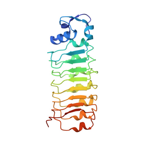
Structure of the lnlB leucine-rich repeats, a domain that triggers host cell invasion by the bacterial pathogen L. monocytogenes.

Marino, M.Braun, L.Cossart, P.Ghosh, P.

(1999) Mol Cell 4: 1063-1072

  • DOI: https://doi.org/10.1016/s1097-2765(00)80234-8
  • Primary Citation Related Structures: 
    1D0B

  • PubMed Abstract: 

    The L. monocytogenes protein lnlB activates phosphoinositide 3-kinase and induces phagocytosis in several mammalian cell types. The 1.86 A resolution X-ray crystal structure of the leucine-rich repeat domain of lnlB that is both necessary and sufficient to induce phagocytosis is presented here. The structure supports a crucial role for calcium in host cell invasion by L. monocytogenes and supplies a rationale for its function. Calciums are bound to the protein in an unusually exposed manner that suggests that the metals may act as a bridge between lnlB and mammalian cell surface receptors. The structure also identifies surfaces on the curved and elongated molecule that may constitute additional interaction sites in forming a bacterial-mammalian signaling complex.


  • Organizational Affiliation
    • Department of Chemistry and Biochemistry, University of California, San Diego, La Jolla 92093-0314, USA.

Macromolecule Content 

  • Total Structure Weight: 23.84 kDa 
  • Atom Count: 1,771 
  • Modeled Residue Count: 207 
  • Deposited Residue Count: 213 
  • Unique protein chains: 1

Macromolecules

Find similar proteins by:|  3D Structure
Entity ID: 1
MoleculeChains  Sequence LengthOrganismDetailsImage
INTERNALIN B213Listeria monocytogenesMutation(s): 0 
UniProt
Find proteins for P0DQD2 (Listeria monocytogenes serovar 1/2a (strain ATCC BAA-679 / EGD-e))
Explore P0DQD2 
Go to UniProtKB:  P0DQD2
Entity Groups
Sequence Clusters30% Identity50% Identity70% Identity90% Identity95% Identity100% Identity
UniProt GroupP0DQD2
Sequence Annotations
Expand
Reference Sequence

Experimental Data & Validation

Experimental Data

  • Method: X-RAY DIFFRACTION
  • Resolution: 1.86 Å
  • R-Value Free:  0.235 (Depositor), 0.235 (DCC) 
  • R-Value Work:  0.207 (Depositor), 0.207 (DCC) 
  • R-Value Observed: 0.210 (Depositor) 
Space Group: P 21 21 21
Unit Cell:
Length ( Å )Angle ( ˚ )
a = 45.41α = 90
b = 56.92β = 90
c = 84.35γ = 90
Software Package:
Software NamePurpose
CCP4model building
SOLVEphasing
SHARPphasing
CNSrefinement
DENZOdata reduction
SCALEPACKdata scaling
CCP4phasing

Structure Validation

View Full Validation Report


Entry History 

Deposition Data

Revision History  (Full details and data files)

  • Version 1.0: 2000-01-07
    Type: Initial release
  • Version 1.1: 2008-04-27
    Changes: Version format compliance
  • Version 1.2: 2011-07-13
    Changes: Version format compliance
  • Version 1.3: 2024-02-07
    Changes: Data collection, Database references, Derived calculations