1CYI | pdb_00001cyi

CYTOCHROME C6


Experimental Data Snapshot

  • Method: X-RAY DIFFRACTION
  • Resolution: 1.90 Å
  • R-Value Work: 
    0.197 (Depositor), 0.200 (DCC) 
  • R-Value Observed: 
    0.197 (Depositor) 

wwPDB Validation   3D Report Full Report


Ligand Structure Quality Assessment 

Created with Raphaël 2.3.0Worse 01 BetterLigand structure goodness of fit to experimental dataBest fitted HECClick on this verticalbar to view details

This is version 2.1 of the entry. See complete history


Literature

The structure of chloroplast cytochrome c6 at 1.9 A resolution: evidence for functional oligomerization.

Kerfeld, C.A.Anwar, H.P.Interrante, R.Merchant, S.Yeates, T.O.

(1995) J Mol Biology 250: 627-647

  • DOI: https://doi.org/10.1006/jmbi.1995.0404
  • Primary Citation of Related Structures:  
    1CYI, 1CYJ

  • PubMed Abstract: 

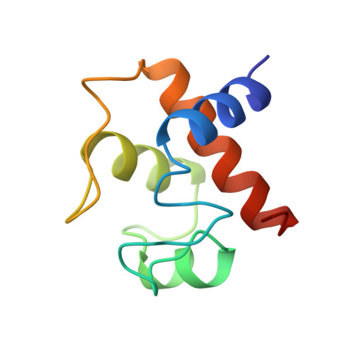
    The molecular structure of cytochrome c6 from the green alga Chlamydomonas reinhardtii has been determined from two crystal forms and refined to 1.9 A resolution. The two crystal forms are likely the result of different levels of post-translational modification of the protein. This is the first report of a high-resolution structure of a chloroplast-derived class I c-type cytochrome. The overall fold is similar to that of other class I c-type cytochromes, consisting of a series of alpha-helices and turns that envelop the heme prosthetic group. There is also a short two-stranded anti-parallel beta-sheet in the vicinity of the methionine axial ligand to the heme; this region of the molecule is formed by the most highly conserved residues in c6-type cytochromes. Although class I c-type cytochromes are assumed to function as monomers, both crystal forms of cytochrome c6 exhibit oligomerization about the heme crevice that is, in part, mediated by the short anti-parallel beta-sheet. The functional significance of this oligomerization is supported by the appearance of similar interfaces in other electron transfer couples, HPLC and light-scattering data, and is furthermore consistent with kinetic data on electron transfer reactions of c6-type cytochromes.


  • Organizational Affiliation

    Molecular Biology Institute, University of California, Los Angeles 90024-1570, USA.


Macromolecules
Find similar proteins by:  (by identity cutoff)  |  3D Structure
Entity ID: 1
MoleculeChains Sequence LengthOrganismDetailsImage
CYTOCHROME C690Chlamydomonas reinhardtiiMutation(s): 0 
UniProt
Find proteins for P08197 (Chlamydomonas reinhardtii)
Explore P08197 
Go to UniProtKB:  P08197
Entity Groups  
Sequence Clusters30% Identity50% Identity70% Identity90% Identity95% Identity100% Identity
UniProt GroupP08197
Sequence Annotations
Expand
  • Reference Sequence
Experimental Data & Validation

Experimental Data

  • Method: X-RAY DIFFRACTION
  • Resolution: 1.90 Å
  • R-Value Work:  0.197 (Depositor), 0.200 (DCC) 
  • R-Value Observed: 0.197 (Depositor) 
Space Group: I 2 3
Unit Cell:
Length ( Å )Angle ( ˚ )
a = 82.6α = 90
b = 82.6β = 90
c = 82.6γ = 90
Software Package:
Software NamePurpose
X-PLORmodel building
X-PLORrefinement
R-AXISdata reduction
MSCdata reduction
X-PLORphasing

Structure Validation

View Full Validation Report


Ligand Structure Quality Assessment 

Created with Raphaël 2.3.0Worse 01 BetterLigand structure goodness of fit to experimental dataBest fitted HECClick on this verticalbar to view details

Entry History 

Deposition Data

Revision History  (Full details and data files)

  • Version 1.0: 1996-01-29
    Type: Initial release
  • Version 1.1: 2008-03-24
    Changes: Version format compliance
  • Version 1.2: 2011-07-13
    Changes: Version format compliance
  • Version 1.3: 2011-11-16
    Changes: Atomic model
  • Version 2.0: 2021-03-10
    Changes: Atomic model, Data collection, Derived calculations, Non-polymer description, Other, Structure summary
  • Version 2.1: 2024-10-30
    Changes: Data collection, Database references, Structure summary