1CR6 | pdb_00001cr6

CRYSTAL STRUCTURE OF MURINE SOLUBLE EPOXIDE HYDROLASE COMPLEXED WITH CPU INHIBITOR


Experimental Data Snapshot

  • Method: X-RAY DIFFRACTION
  • Resolution: 2.80 Å
  • R-Value Free: 
    0.312 (Depositor) 
  • R-Value Work: 
    0.201 (Depositor) 
  • R-Value Observed: 
    0.252 (Depositor) 

wwPDB Validation 3D Report Full Report

Validation slider image for 1CR6

This is version 1.3 of the entry. See complete history

Literature

Detoxification of environmental mutagens and carcinogens: structure, mechanism, and evolution of liver epoxide hydrolase.

Argiriadi, M.A.Morisseau, C.Hammock, B.D.Christianson, D.W.

(1999) Proc Natl Acad Sci U S A 96: 10637-10642

  • DOI: https://doi.org/10.1073/pnas.96.19.10637
  • Primary Citation Related Structures: 
    1CQZ, 1CR6

  • PubMed Abstract: 

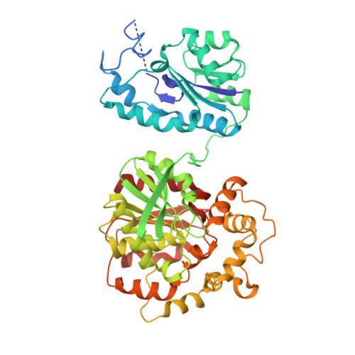
    The crystal structure of recombinant murine liver cytosolic epoxide hydrolase (EC 3.3.2.3) has been determined at 2.8-A resolution. The binding of a nanomolar affinity inhibitor confirms the active site location in the C-terminal domain; this domain is similar to that of haloalkane dehalogenase and shares the alpha/beta hydrolase fold. A structure-based mechanism is proposed that illuminates the unique chemical strategy for the activation of endogenous and man-made epoxide substrates for hydrolysis and detoxification. Surprisingly, a vestigial active site is found in the N-terminal domain similar to that of another enzyme of halocarbon metabolism, haloacid dehalogenase. Although the vestigial active site does not participate in epoxide hydrolysis, the vestigial domain plays a critical structural role by stabilizing the dimer in a distinctive domain-swapped architecture. Given the genetic and structural relationships among these enzymes of xenobiotic metabolism, a structure-based evolutionary sequence is postulated.


  • Organizational Affiliation
    • Roy and Diana Vagelos Laboratories, Department of Chemistry, University of Pennsylvania, Philadelphia, PA 19104-6323, USA.

Macromolecule Content 

  • Total Structure Weight: 125.68 kDa 
  • Atom Count: 8,237 
  • Modeled Residue Count: 1,028 
  • Deposited Residue Count: 1,108 
  • Unique protein chains: 1

Macromolecules

Find similar proteins by:|  3D Structure
Entity ID: 1
MoleculeChains  Sequence LengthOrganismDetailsImage
EPOXIDE HYDROLASE
A, B
554Mus musculusMutation(s): 0 
EC: 3.3.2.3 (PDB Primary Data), 3.1.3.76 (UniProt), 3.3.2.10 (UniProt)
UniProt
Find proteins for P34914 (Mus musculus)
Explore P34914 
Go to UniProtKB:  P34914
Entity Groups
Sequence Clusters30% Identity50% Identity70% Identity90% Identity95% Identity100% Identity
UniProt GroupP34914
Sequence Annotations
Expand
Reference Sequence

Small Molecules

Ligands 1 Unique
IDChains Name / Formula / InChI Key2D Diagram3D Interactions
CPU

Query on CPU


Download:Ideal Coordinates CCD File
C [auth A],
D [auth B]
N-CYCLOHEXYL-N'-(PROPYL)PHENYL UREA
C16 H24 N2 O
HBTZVNKXMFGOOJ-UHFFFAOYSA-N

Experimental Data & Validation

Experimental Data

  • Method: X-RAY DIFFRACTION
  • Resolution: 2.80 Å
  • R-Value Free:  0.312 (Depositor) 
  • R-Value Work:  0.201 (Depositor) 
  • R-Value Observed: 0.252 (Depositor) 
Space Group: P 21 21 2
Unit Cell:
Length ( Å )Angle ( ˚ )
a = 151.9α = 90
b = 143β = 90
c = 60γ = 90
Software Package:
Software NamePurpose
X-PLORmodel building
X-PLORrefinement
DENZOdata reduction
SCALEPACKdata scaling
X-PLORphasing

Structure Validation

View Full Validation Report


Entry History 

Deposition Data

Revision History  (Full details and data files)

  • Version 1.0: 1999-11-19
    Type: Initial release
  • Version 1.1: 2008-04-27
    Changes: Version format compliance
  • Version 1.2: 2011-07-13
    Changes: Version format compliance
  • Version 1.3: 2024-02-07
    Changes: Data collection, Database references, Derived calculations