1CQ1 | pdb_00001cq1

Soluble Quinoprotein Glucose Dehydrogenase from Acinetobacter Calcoaceticus in Complex with PQQH2 and Glucose


Experimental Data Snapshot

  • Method: X-RAY DIFFRACTION
  • Resolution: 1.90 Å
  • R-Value Free: 
    0.226 (Depositor) 
  • R-Value Work: 
    0.190 (Depositor), 0.186 (DCC) 
  • R-Value Observed: 
    0.190 (Depositor) 

wwPDB Validation 3D Report Full Report

Validation slider image for 1CQ1

Ligand Structure Quality Assessment 


This is version 1.7 of the entry. See complete history

Literature

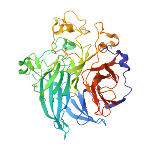
Structure and mechanism of soluble quinoprotein glucose dehydrogenase.

Oubrie, A.Rozeboom, H.J.Kalk, K.H.Olsthoorn, A.J.Duine, J.A.Dijkstra, B.W.

(1999) EMBO J 18: 5187-5194

  • DOI: https://doi.org/10.1093/emboj/18.19.5187
  • Primary Citation Related Structures: 
    1C9U, 1CQ1

  • PubMed Abstract: 

    Soluble glucose dehydrogenase (s-GDH; EC 1.1.99.17) is a classical quinoprotein which requires the cofactor pyrroloquinoline quinone (PQQ) to oxidize glucose to gluconolactone. The reaction mechanism of PQQ-dependent enzymes has remained controversial due to the absence of comprehensive structural data. We have determined the X-ray structure of s-GDH with the cofactor at 2.2 A resolution, and of a complex with reduced PQQ and glucose at 1.9 A resolution. These structures reveal the active site of s-GDH, and show for the first time how a functionally bound substrate interacts with the cofactor in a PQQ-dependent enzyme. Twenty years after the discovery of PQQ, our results finally provide conclusive evidence for a reaction mechanism comprising general base-catalyzed hydride transfer, rather than the generally accepted covalent addition-elimination mechanism. Thus, PQQ-dependent enzymes use a mechanism similar to that of nicotinamide- and flavin-dependent oxidoreductases.


  • Organizational Affiliation
    • Laboratory of Biophysical Chemistry and BIOSON Research Institute, University of Groningen, Nijenborgh 4, 9747 AG Groningen.

Macromolecule Content 

  • Total Structure Weight: 101.85 kDa 
  • Atom Count: 7,629 
  • Modeled Residue Count: 890 
  • Deposited Residue Count: 908 
  • Unique protein chains: 1

Macromolecules

Find similar proteins by:|  3D Structure
Entity ID: 1
MoleculeChains  Sequence LengthOrganismDetailsImage
SOLUBLE QUINOPROTEIN GLUCOSE DEHYDROGENASE
A, B
454Acinetobacter calcoaceticusMutation(s): 0 
EC: 1.1.5.2
UniProt
Find proteins for P13650 (Acinetobacter calcoaceticus)
Explore P13650 
Go to UniProtKB:  P13650
Entity Groups
Sequence Clusters30% Identity50% Identity70% Identity90% Identity95% Identity100% Identity
UniProt GroupP13650
Sequence Annotations
Expand
Reference Sequence

Small Molecules

Ligands 3 Unique
IDChains Name / Formula / InChI Key2D Diagram3D Interactions
PQQ

Query on PQQ


Download:Ideal Coordinates CCD File
G [auth A],
L [auth B]
PYRROLOQUINOLINE QUINONE
C14 H6 N2 O8
MMXZSJMASHPLLR-UHFFFAOYSA-N
BGC

Query on BGC


Download:Ideal Coordinates CCD File
C [auth A],
H [auth B]
beta-D-glucopyranose
C6 H12 O6
WQZGKKKJIJFFOK-VFUOTHLCSA-N
CA

Query on CA


Download:Ideal Coordinates CCD File
D [auth A]
E [auth A]
F [auth A]
I [auth B]
J [auth B]
D [auth A],
E [auth A],
F [auth A],
I [auth B],
J [auth B],
K [auth B]
CALCIUM ION
Ca
BHPQYMZQTOCNFJ-UHFFFAOYSA-N

Experimental Data & Validation

Experimental Data

  • Method: X-RAY DIFFRACTION
  • Resolution: 1.90 Å
  • R-Value Free:  0.226 (Depositor) 
  • R-Value Work:  0.190 (Depositor), 0.186 (DCC) 
  • R-Value Observed: 0.190 (Depositor) 
Space Group: P 1 21 1
Unit Cell:
Length ( Å )Angle ( ˚ )
a = 60.899α = 90
b = 91.972β = 104.62
c = 85.747γ = 90
Software Package:
Software NamePurpose
X-PLORrefinement
SCALEPACKdata scaling
REFMACrefinement
X-PLORmodel building
DENZOdata reduction
X-PLORphasing

Structure Validation

View Full Validation Report


Ligand Structure Quality Assessment 


Entry History 

Deposition Data

Revision History  (Full details and data files)

  • Version 1.0: 2000-02-04
    Type: Initial release
  • Version 1.1: 2007-10-21
    Changes: Version format compliance
  • Version 1.2: 2011-07-13
    Changes: Version format compliance
  • Version 1.3: 2017-10-04
    Changes: Refinement description
  • Version 1.4: 2019-07-24
    Changes: Data collection, Refinement description
  • Version 1.5: 2019-08-14
    Changes: Data collection
  • Version 1.6: 2020-07-29
    Type: Remediation
    Reason: Carbohydrate remediation
    Changes: Data collection, Derived calculations, Structure summary
  • Version 1.7: 2024-10-30
    Changes: Data collection, Database references, Structure summary