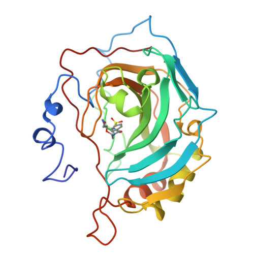
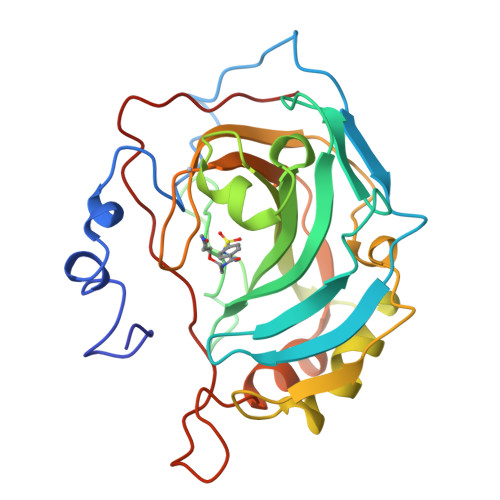
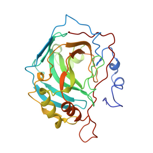
1CNX

SECONDARY INTERACTIONS SIGNIFICANTLY REMOVED FROM THE SULFONAMIDE BINDING POCKET OF CARBONIC ANHYDRASE II INFLUENCE BINDING CONSTANTS


Experimental Data Snapshot

  • Method: X-RAY DIFFRACTION
  • Resolution: 1.90 Å
  • R-Value Observed: 0.174 

wwPDB Validation   3D Report Full Report


This is version 1.3 of the entry. See complete history


Literature

Secondary interactions significantly removed from the sulfonamide binding pocket of carbonic anhydrase II influence inhibitor binding constants.

Boriack, P.A.Christianson, D.W.Kingery-Wood, J.Whitesides, G.M.

(1995) J Med Chem 38: 2286-2291

  • DOI: https://doi.org/10.1021/jm00013a004
  • Primary Citation of Related Structures:  
    1CNW, 1CNX, 1CNY

  • PubMed Abstract: 

    A series of competitive inhibitors of carbonic anhydrase II (CAII; EC 4.2.1.1) that consists of oligo(ethylene glycol) units attached to p-benzenesulfonamides with pendant amino acids, H2NSO2C6H4CONHCH2CH2OCH2CH2OCH2CH2NHCOCHRNH3+, have been synthesized and examined using competitive fluorescence assays. Three of the strongest inhibitors, designated EG3NH3+, EG3GlyNH3+, and EG3PheNH3+, have been studied by X-ray crystallographic methods at limiting resolutions of 1.9, 2.0, and 2.3 A, respectively. The sulfonamide-zinc binding modes and the association of the ethylene glycol linkers to the hydrophobic patch of the active site are similar in all three inhibitors. Differences in the values of Kd are therefore not due to differences in zinc coordination or to differences in the modes of enzyme-glycol association but instead appear to arise from interaction of the pendant amino acids with the surface of the protein. These pendant groups are, however, not sufficiently ordered to be visible in electron density maps. Thus, structural variations of inhibitors at locations distant from the primary binding (i.e., the sulfonamide group) site affect the overall binding affinities of inhibitors (e.g., Kd (EG3PheNH3+) = 14 nM as compared with Kd (EG3GluNH3+) = 100 nM).


  • Organizational Affiliation

    Department of Chemistry, University of Pennsylvania, Philadelphia 19104-6323, USA.


Macromolecules
Find similar proteins by:  (by identity cutoff)  |  3D Structure
Entity ID: 1
MoleculeChains Sequence LengthOrganismDetailsImage
CARBONIC ANHYDRASE II260Homo sapiensMutation(s): 0 
EC: 4.2.1.1 (PDB Primary Data), 4.2.1.69 (UniProt)
UniProt & NIH Common Fund Data Resources
Find proteins for P00918 (Homo sapiens)
Explore P00918 
Go to UniProtKB:  P00918
PHAROS:  P00918
GTEx:  ENSG00000104267 
Entity Groups  
Sequence Clusters30% Identity50% Identity70% Identity90% Identity95% Identity100% Identity
UniProt GroupP00918
Sequence Annotations
Expand
  • Reference Sequence
Small Molecules
Ligands 3 Unique
IDChains Name / Formula / InChI Key2D Diagram3D Interactions
EG2
Query on EG2

Download Ideal Coordinates CCD File 
D [auth A]AMINODI(ETHYLOXY)ETHYLAMINOCARBONYLBENZENESULFONAMIDE
C13 H21 N3 O5 S
QGGYVKRXFQLYQD-UHFFFAOYSA-N
HG
Query on HG

Download Ideal Coordinates CCD File 
C [auth A]MERCURY (II) ION
Hg
BQPIGGFYSBELGY-UHFFFAOYSA-N
ZN
Query on ZN

Download Ideal Coordinates CCD File 
B [auth A]ZINC ION
Zn
PTFCDOFLOPIGGS-UHFFFAOYSA-N
Binding Affinity Annotations 
IDSourceBinding Affinity
EG2 PDBBind:  1CNX Kd: 43 (nM) from 1 assay(s)
Experimental Data & Validation

Experimental Data

  • Method: X-RAY DIFFRACTION
  • Resolution: 1.90 Å
  • R-Value Observed: 0.174 
  • Space Group: P 1 21 1
Unit Cell:
Length ( Å )Angle ( ˚ )
a = 42.7α = 90
b = 41.7β = 104.6
c = 73γ = 90
Software Package:
Software NamePurpose
PROLSQrefinement

Structure Validation

View Full Validation Report


Entry History 

Deposition Data

Revision History  (Full details and data files)

  • Version 1.0: 1995-11-14
    Type: Initial release
  • Version 1.1: 2008-03-24
    Changes: Version format compliance
  • Version 1.2: 2011-07-13
    Changes: Version format compliance
  • Version 1.3: 2024-02-07
    Changes: Data collection, Database references, Derived calculations, Other