1CLO | pdb_00001clo

ANTI-CARCINOEMBRYONIC ANTIGEN MONOCLONAL ANTIBODY A5B7


Experimental Data Snapshot

  • Method: X-RAY DIFFRACTION
  • Resolution: 2.10 Å
  • R-Value Work: 
    0.212 (Depositor), 0.190 (DCC) 
  • R-Value Observed: 
    0.212 (Depositor) 

wwPDB Validation 3D Report Full Report

Validation slider image for 1CLO

This is version 1.3 of the entry. See complete history

Literature

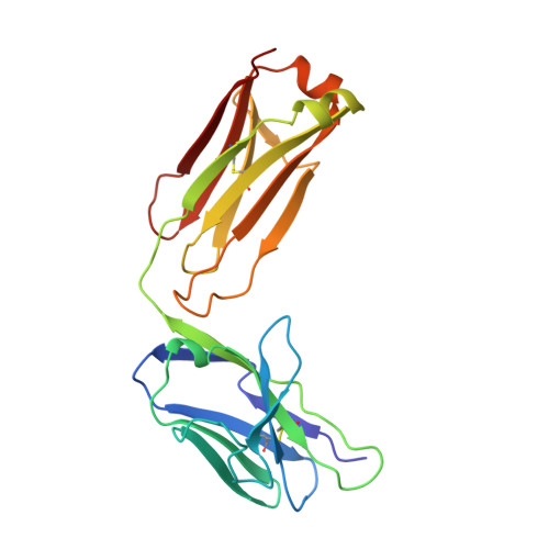
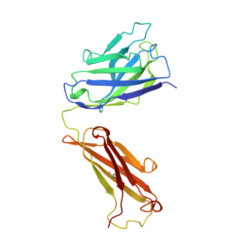
VL:VH domain rotations in engineered antibodies: crystal structures of the Fab fragments from two murine antitumor antibodies and their engineered human constructs.

Banfield, M.J.King, D.J.Mountain, A.Brady, R.L.

(1997) Proteins 29: 161-171

  • DOI: https://doi.org/10.1002/(sici)1097-0134(199710)29:2<161::aid-prot4>3.0.co;2-g
  • Primary Citation Related Structures: 
    1AD0, 1AD9, 1AE6, 1CLO

  • PubMed Abstract: 

    The crystal structures of two pairs of Fab fragments have been determined. The pairs comprise both a murine and an engineered human form, each derived from the antitumor antibodies A5B7 and CTM01. Although antigen specificity is maintained within the pairs, antigen affinity varies. A comparison of the hypervariable loops for each pair of antibodies shows their structure has been well maintained in grafting, supporting the canonical loop model. Detailed structural analysis of the binding sites and domain arrangements for these antibodies suggests the differences in antigen affinity observed are likely to be due to inherent flexibility of the hypervariable loops and movements at the VL:VH domain interface. The four structures provide the first opportunity to study in detail the effects of protein engineering on specific antibodies.


  • Organizational Affiliation
    • Molecular Recognition Centre, University of Bristol School of Medical Sciences, United Kingdom.

Macromolecule Content 

  • Total Structure Weight: 47.31 kDa 
  • Atom Count: 3,727 
  • Modeled Residue Count: 435 
  • Deposited Residue Count: 435 
  • Unique protein chains: 2

Macromolecules

Find similar proteins by:|  3D Structure
Entity ID: 1
MoleculeChains  Sequence LengthOrganismDetailsImage
A5B7 MONOCLONAL ANTIBODYA [auth L]213Mus musculusMutation(s): 0 
Find similar proteins by:|  3D Structure
Entity ID: 2
MoleculeChains  Sequence LengthOrganismDetailsImage
A5B7 MONOCLONAL ANTIBODYB [auth H]222Mus musculusMutation(s): 0 
UniProt
Find proteins for P01868 (Mus musculus)
Explore P01868 
Go to UniProtKB:  P01868
Entity Groups
Sequence Clusters30% Identity50% Identity70% Identity90% Identity95% Identity100% Identity
UniProt GroupP01868
Sequence Annotations
Expand
Reference Sequence

Experimental Data & Validation

Experimental Data

  • Method: X-RAY DIFFRACTION
  • Resolution: 2.10 Å
  • R-Value Work:  0.212 (Depositor), 0.190 (DCC) 
  • R-Value Observed: 0.212 (Depositor) 
Space Group: P 1 21 1
Unit Cell:
Length ( Å )Angle ( ˚ )
a = 38.89α = 90
b = 79.68β = 104.91
c = 68.52γ = 90
Software Package:
Software NamePurpose
DENZOdata reduction
SCALEPACKdata scaling
AMoREphasing
X-PLORrefinement

Structure Validation

View Full Validation Report


Entry History 

Deposition Data

Revision History  (Full details and data files)

  • Version 1.0: 1997-05-15
    Type: Initial release
  • Version 1.1: 2008-03-24
    Changes: Version format compliance
  • Version 1.2: 2011-07-13
    Changes: Version format compliance
  • Version 1.3: 2024-10-30
    Changes: Data collection, Database references, Structure summary