1CL7 | pdb_00001cl7

ANTI HIV1 PROTEASE FAB


Experimental Data Snapshot

  • Method: X-RAY DIFFRACTION
  • Resolution: 3.00 Å
  • R-Value Free: 
    0.280 (Depositor) 
  • R-Value Work: 
    0.183 (Depositor) 

Starting Model: experimental
View more details

wwPDB Validation 3D Report Full Report

Validation slider image for 1CL7

This is version 1.4 of the entry. See complete history

Literature

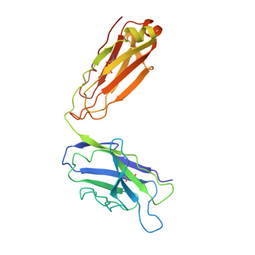
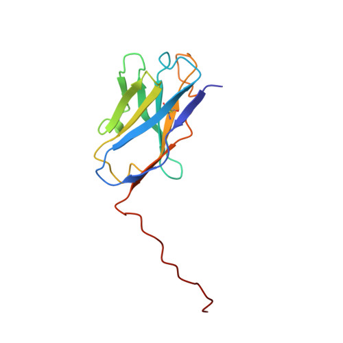
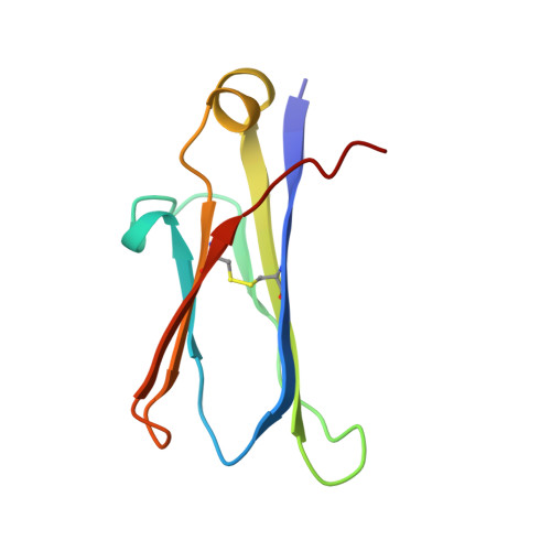
Inhibition of the HIV-1 and HIV-2 proteases by a monoclonal antibody.

Lescar, J.Brynda, J.Rezacova, P.Stouracova, R.Riottot, M.M.Chitarra, V.Fabry, M.Horejsi, M.Sedlacek, J.Bentley, G.A.

(1999) Protein Sci 8: 2686-2696

  • DOI: https://doi.org/10.1110/ps.8.12.2686
  • Primary Citation Related Structures: 
    1CL7

  • PubMed Abstract: 

    The monoclonal antibody 1696, directed against the HIV-1 protease, displays strong inhibitory effects toward the catalytic activity of the enzyme of both the HIV-1 and HIV-2 isolates. This antibody cross-reacts with peptides that include the N-terminus of the enzyme, a region that is well conserved in sequence among different viral strains and which, furthermore, is crucial for homodimerization to the active enzymatic form. This observation, as well as antigen-binding studies in the presence of an active site inhibitor, suggest that 1696 inhibits the HIV protease by destabilizing its active homodimeric form. To characterize further how the antibody 1696 inhibits the HIV-1 and HIV-2 proteases, we have solved the crystal structure of its Fab fragment by molecular replacement and refined it at 3.0 A resolution. The antigen binding site has a deep cavity at its center, which is lined mainly by acidic and hydrophobic residues, and is large enough to accommodate several antigen residues. The structure of the Fab 1696 could form a starting basis for the design of alternative HIV protease-inhibiting molecules of broad specificity.


  • Organizational Affiliation
    • Unité d'Immunologie Structurale (URA 1961 CNRS), Département d'Immunologie, Institut Pasteur, Paris, France.

Macromolecule Content 

  • Total Structure Weight: 47.66 kDa 
  • Atom Count: 3,355 
  • Modeled Residue Count: 433 
  • Deposited Residue Count: 433 
  • Unique protein chains: 3

Macromolecules

Find similar proteins by:|  3D Structure
Entity ID: 1
MoleculeChains  Sequence LengthOrganismDetailsImage
PROTEIN (IGG1 ANTIBODY 1696 (light chain))A [auth L]216Mus musculusMutation(s): 0 
Find similar proteins by:|  3D Structure
Entity ID: 2
MoleculeChains  Sequence LengthOrganismDetailsImage
PROTEIN (IGG1 ANTIBODY 1696 (variable heavy chain))B [auth H]135Mus musculusMutation(s): 0 
Find similar proteins by:|  3D Structure
Entity ID: 3
MoleculeChains  Sequence LengthOrganismDetailsImage
PROTEIN (IGG1 ANTIBODY 1696 (constant heavy chain))C [auth I]82Mus musculusMutation(s): 0 
UniProt
Find proteins for P01869 (Mus musculus)
Explore P01869 
Go to UniProtKB:  P01869
Entity Groups
Sequence Clusters30% Identity50% Identity70% Identity90% Identity95% Identity100% Identity
UniProt GroupP01869
Sequence Annotations
Expand
Reference Sequence

Experimental Data & Validation

Experimental Data

  • Method: X-RAY DIFFRACTION
  • Resolution: 3.00 Å
  • R-Value Free:  0.280 (Depositor) 
  • R-Value Work:  0.183 (Depositor) 
Space Group: P 21 21 21
Unit Cell:
Length ( Å )Angle ( ˚ )
a = 46.4α = 90
b = 58.8β = 90
c = 143γ = 90
Software Package:
Software NamePurpose
DENZOdata reduction
SCALEPACKdata scaling
AMoREphasing
X-PLORrefinement

Structure Validation

View Full Validation Report


Entry History 

Deposition Data

Revision History  (Full details and data files)

  • Version 1.0: 2000-01-12
    Type: Initial release
  • Version 1.1: 2008-04-26
    Changes: Version format compliance
  • Version 1.2: 2011-07-13
    Changes: Version format compliance
  • Version 1.3: 2023-08-09
    Changes: Data collection, Database references, Refinement description
  • Version 1.4: 2024-11-13
    Changes: Structure summary