1CID | pdb_00001cid

CRYSTAL STRUCTURE OF DOMAINS 3 & 4 OF RAT CD4 AND THEIR RELATIONSHIP TO THE NH2-TERMINAL DOMAINS


Experimental Data Snapshot

  • Method: X-RAY DIFFRACTION
  • Resolution: 2.80 Å

wwPDB Validation 3D Report Full Report

Validation slider image for 1CID

This is version 2.1 of the entry. See complete history

Literature

Crystal structure of domains 3 and 4 of rat CD4: relation to the NH2-terminal domains.

Brady, R.L.Dodson, E.J.Dodson, G.G.Lange, G.Davis, S.J.Williams, A.F.Barclay, A.N.

(1993) Science 260: 979-983

  • DOI: https://doi.org/10.1126/science.8493535
  • Primary Citation Related Structures: 
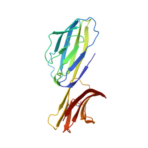
    1CID

  • PubMed Abstract: 

    The CD4 antigen is a membrane glycoprotein of T lymphocytes that interacts with major histocompatibility complex class II antigens and is also a receptor for the human immunodeficiency virus. the extracellular portion of CD4 is predicted to fold into four immunoglobulin-like domains. The crystal structure of the third and fourth domains of rat CD4 was solved at 2.8 angstrom resolution and shows that both domains have immunoglobulin folds. Domain 3, however, lacks the disulfide between the beta sheets; this results in an expansion of the domain. There is a difference of 30 degrees in the orientation between domains 3 and 4 when compared with domains 1 and 2. The two CD4 fragment structures provide a basis from which models of the overall receptor can be proposed. These models suggest an extended structure comprising two rigid portions joined by a short and possibly flexible linker region.


  • Organizational Affiliation
    • Department of Chemistry, University of York, United Kingdom.

Macromolecule Content 

  • Total Structure Weight: 19.8 kDa 
  • Atom Count: 1,396 
  • Modeled Residue Count: 177 
  • Deposited Residue Count: 177 
  • Unique protein chains: 1

Macromolecules

Find similar proteins by:|  3D Structure
Entity ID: 1
MoleculeChains  Sequence LengthOrganismDetailsImage
T CELL SURFACE GLYCOPROTEIN CD4177Rattus norvegicusMutation(s): 0 
UniProt
Find proteins for P05540 (Rattus norvegicus)
Explore P05540 
Go to UniProtKB:  P05540
Entity Groups
Sequence Clusters30% Identity50% Identity70% Identity90% Identity95% Identity100% Identity
UniProt GroupP05540
Sequence Annotations
Expand
Reference Sequence

Small Molecules

Ligands 1 Unique
IDChains Name / Formula / InChI Key2D Diagram3D Interactions
SO4

Query on SO4


Download:Ideal Coordinates CCD File
B [auth A]SULFATE ION
O4 S
QAOWNCQODCNURD-UHFFFAOYSA-L

Experimental Data & Validation

Experimental Data

  • Method: X-RAY DIFFRACTION
  • Resolution: 2.80 Å
Space Group: P 31 2 1
Unit Cell:
Length ( Å )Angle ( ˚ )
a = 77.8α = 90
b = 77.8β = 90
c = 82.5γ = 120
Software Package:
Software NamePurpose
PROLSQrefinement

Structure Validation

View Full Validation Report


Entry History 

Deposition Data

Revision History  (Full details and data files)

  • Version 1.0: 1993-07-15
    Type: Initial release
  • Version 1.1: 2008-03-24
    Changes: Version format compliance
  • Version 1.2: 2011-07-13
    Changes: Version format compliance
  • Version 2.0: 2024-06-05
    Changes: Atomic model, Data collection, Database references, Other
  • Version 2.1: 2024-11-20
    Changes: Advisory, Derived calculations, Structure summary