1CA6 | pdb_00001ca6

INTERCALATION SITE OF HYPERTHERMOPHILE CHROMOSOMAL PROTEIN SSO7D/SAC7D BOUND TO DNA


Experimental Data Snapshot

  • Method: X-RAY DIFFRACTION
  • Resolution: 2.20 Å
  • R-Value Free: 
    0.257 (Depositor) 
  • R-Value Work: 
    0.197 (Depositor) 

Starting Model: experimental
View more details

wwPDB Validation 3D Report Full Report

Validation slider image for 1CA6

This is version 1.6 of the entry. See complete history

Literature

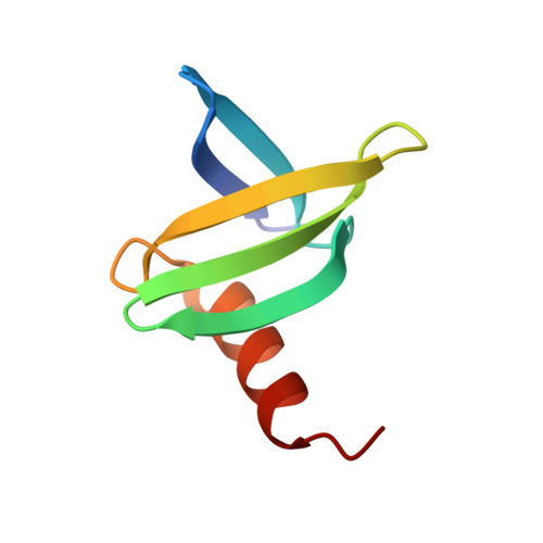
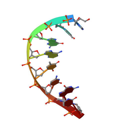
Crystal structures of the chromosomal proteins Sso7d/Sac7d bound to DNA containing T-G mismatched base-pairs

Su, S.Gao, Y.-G.Robinson, H.Liaw, Y.C.Edmondson, S.P.Shriver, J.W.Wang, A.H.-J.

(2000) J Mol Biology 303: 395-403

  • DOI: https://doi.org/10.1006/jmbi.2000.4112
  • Primary Citation Related Structures: 
    1C8C, 1CA5, 1CA6

  • PubMed Abstract: 

    Sso7d and Sac7d are two small chromatin proteins from the hyperthermophilic archaeabacterium Sulfolobus solfataricus and Sulfolobus acidocaldarius, respectively. The crystal structures of Sso7d-GTGATCGC, Sac7d-GTGATCGC and Sac7d-GTGATCAC have been determined and refined at 1.45 A, 2.2 A and 2.2 A, respectively, to investigate the DNA binding property of Sso7d/Sac7d in the presence of a T-G mismatch base-pair. Detailed structural analysis revealed that the intercalation site includes the T-G mismatch base-pair and Sso7d/Sac7d bind to that mismatch base-pair in a manner similar to regular DNA. In the Sso7d-GTGATCGC complex, a new inter-strand hydrogen bond between T2O4 and C14N4 is formed and well-order bridging water molecules are found. The results suggest that the less stable DNA stacking site involving a T-G mismatch may be a preferred site for protein side-chain intercalation.


  • Organizational Affiliation
    • Center for Biophysics and COmputational BIology, University of Illinois at Urbana-Champaign, 61801, USA.

Macromolecule Content 

  • Total Structure Weight: 12.51 kDa 
  • Atom Count: 929 
  • Modeled Residue Count: 82 
  • Deposited Residue Count: 82 
  • Unique protein chains: 1
  • Unique nucleic acid chains: 1

Macromolecules


Find similar proteins by:|  3D Structure
Entity ID: 2
MoleculeChains  Sequence LengthOrganismDetailsImage
CHROMOSOMAL PROTEIN SAC7DC [auth A]66Sulfolobus acidocaldariusMutation(s): 0 
UniProt
Find proteins for P13123 (Sulfolobus acidocaldarius (strain ATCC 33909 / DSM 639 / JCM 8929 / NBRC 15157 / NCIMB 11770))
Explore P13123 
Go to UniProtKB:  P13123
Entity Groups
Sequence Clusters30% Identity50% Identity70% Identity90% Identity95% Identity100% Identity
UniProt GroupP13123
Sequence Annotations
Expand
Reference Sequence
Find similar nucleic acids by:  Sequence
Entity ID: 1
MoleculeChains LengthOrganismImage
5'-D(*GP*TP*GP*AP*TP*CP*GP*C)-3'A [auth B],
B [auth C]
8synthetic construct
Sequence Annotations
Expand
Reference Sequence

Experimental Data & Validation

Experimental Data

  • Method: X-RAY DIFFRACTION
  • Resolution: 2.20 Å
  • R-Value Free:  0.257 (Depositor) 
  • R-Value Work:  0.197 (Depositor) 
Space Group: P 21 21 21
Unit Cell:
Length ( Å )Angle ( ˚ )
a = 50.47α = 90
b = 75.89β = 90
c = 35γ = 90
Software Package:
Software NamePurpose
X-PLORmodel building
X-PLORrefinement
X-PLORphasing

Structure Validation

View Full Validation Report


Entry History 

Deposition Data

Revision History  (Full details and data files)

  • Version 1.0: 2000-02-23
    Type: Initial release
  • Version 1.1: 2008-04-26
    Changes: Version format compliance
  • Version 1.2: 2011-07-13
    Changes: Version format compliance
  • Version 1.3: 2011-12-28
    Changes: Advisory
  • Version 1.4: 2017-10-04
    Changes: Refinement description
  • Version 1.5: 2019-11-06
    Changes: Data collection, Database references, Source and taxonomy
  • Version 1.6: 2023-08-09
    Changes: Data collection, Database references, Refinement description