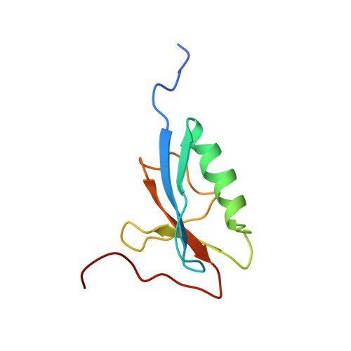
Structure of the CAD domain of caspase-activated DNase and interaction with the CAD domain of its inhibitor.
Uegaki, K., Otomo, T., Sakahira, H., Shimizu, M., Yumoto, N., Kyogoku, Y., Nagata, S., Yamazaki, T.(2000) J Mol Biology 297: 1121-1128
- PubMed: 10764577 Search on PubMed
- DOI: https://doi.org/10.1006/jmbi.2000.3643
- Primary Citation Related Structures:
1C9F - PubMed Abstract:
Caspase-activated DNase (CAD), which causes a genome fragmentation at the final stage of apoptosis, is a protein of about 40 kDa and exists as a complex form with the inhibitor ICAD in living cells. There is sequence homology of about 80 amino acid residues at the N termini of CAD and ICAD (called the CAD domain). Here, we report the three-dimensional structure of the CAD domain of CAD determined by multi-dimensional NMR spectroscopy and the property of CAD domains investigated by a surface plasmon resonance experiment. The CAD domain of CAD is an independently folded domain composed of one alpha-helix and five beta-strands forming a single sheet. The overall structure is categorized in the ubiquitin superfold. This domain can bind strongly to the isolated CAD domain of ICAD (dissociation constant: 5.48(+/-0.003)x10(-8) M). It suggests the function of the CAD domains in the CAD-ICAD system, that the protein-protein interaction through the CAD domains plays an important role in the inhibition of CAD DNase activity and in the correct folding of CAD. On the basis of structural comparison with other protein complexes containing the ubiquitin superfold, the interaction mode of the CAD domains is proposed.
- Osaka National Research Institute, AIST, 1-8-31 Midorigaoka, Ikeda, Osaka, 563-8577, Japan.
Organizational Affiliation: