1C54 | pdb_00001c54

SOLUTION STRUCTURE OF RIBONUCLEASE SA


Experimental Data Snapshot

  • Method: SOLUTION NMR
  • Conformers Calculated: 40 
  • Conformers Submitted: 20 
  • Selection Criteria: LOWEST ENERGY AND REPRESENTATIVE OF DIFFERENT CONFORMERS 

wwPDB Validation 3D Report Full Report

Validation slider image for 1C54

This is version 1.5 of the entry. See complete history

Literature

Solution structure and dynamics of ribonuclease Sa.

Laurents, D.Perez-Canadillas, J.M.Santoro, J.Rico, M.Schell, D.Pace, C.N.Bruix, M.

(2001) Proteins 44: 200-211

  • DOI: https://doi.org/10.1002/prot.1085
  • Primary Citation Related Structures: 
    1C54

  • PubMed Abstract: 

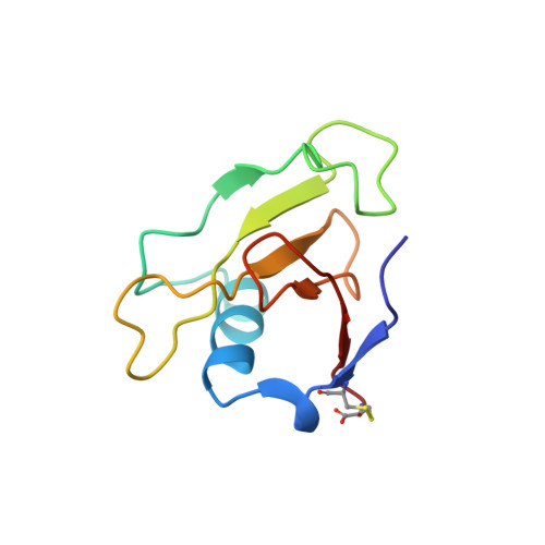
    We have used NMR methods to characterize the structure and dynamics of ribonuclease Sa in solution. The solution structure of RNase Sa was obtained using the distance constraints provided by 2,276 NOEs and the C6-C96 disulfide bond. The 40 resulting structures are well determined; their mean pairwise RMSD is 0.76 A (backbone) and 1.26 A (heavy atoms). The solution structures are similar to previously determined crystal structures, especially in the secondary structure, but exhibit new features: the loop composed of Pro 45 to Ser 48 adopts distinct conformations and the rings of tyrosines 51, 52, and 55 have reduced flipping rates. Amide protons with greatly reduced exchange rates are found predominantly in interior beta-strands and the alpha-helix, but also in the external 3/10 helix and edge beta-strand linked by the disulfide bond. Analysis of (15)N relaxation experiments (R1, R2, and NOE) at 600 MHz revealed five segments, consisting of residues 1-5, 28-31, 46-50, 60-65, 74-77, retaining flexibility in solution. The change in conformation entropy for RNase SA folding is smaller than previously believed, since the native protein is more flexible in solution than in a crystal.


  • Organizational Affiliation
    • Instituto de Estructura de la Materia, CSIC, Madrid, Spain.

Macromolecule Content 

  • Total Structure Weight: 10.58 kDa 
  • Atom Count: 746 
  • Modeled Residue Count: 96 
  • Deposited Residue Count: 96 
  • Unique protein chains: 1

Macromolecules

Find similar proteins by:|  3D Structure
Entity ID: 1
MoleculeChains  Sequence LengthOrganismDetailsImage
RIBONUCLEASE SA96Kitasatospora aureofaciensMutation(s): 0 
EC: 3.1.27.3 (PDB Primary Data), 4.6.1.24 (UniProt)
UniProt
Find proteins for P05798 (Kitasatospora aureofaciens)
Explore P05798 
Go to UniProtKB:  P05798
Entity Groups
Sequence Clusters30% Identity50% Identity70% Identity90% Identity95% Identity100% Identity
UniProt GroupP05798
Sequence Annotations
Expand
Reference Sequence

Experimental Data & Validation

Experimental Data

  • Method: SOLUTION NMR
  • Conformers Calculated: 40 
  • Conformers Submitted: 20 
  • Selection Criteria: LOWEST ENERGY AND REPRESENTATIVE OF DIFFERENT CONFORMERS 

Structure Validation

View Full Validation Report


Entry History 

Deposition Data

Revision History  (Full details and data files)

  • Version 1.0: 2001-11-28
    Type: Initial release
  • Version 1.1: 2008-04-26
    Changes: Version format compliance
  • Version 1.2: 2011-07-13
    Changes: Version format compliance
  • Version 1.3: 2022-02-16
    Changes: Data collection, Database references, Derived calculations
  • Version 1.4: 2023-12-27
    Changes: Data collection
  • Version 1.5: 2024-10-09
    Changes: Structure summary