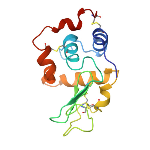
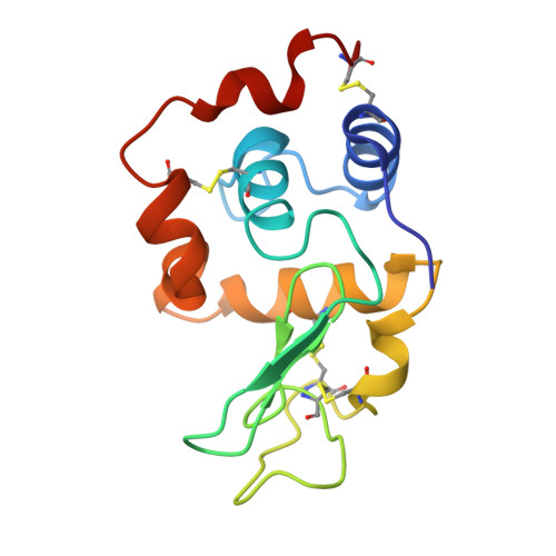
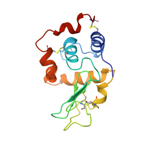
1C46

MUTANT HUMAN LYSOZYME WITH FOREIGN N-TERMINAL RESIDUES


Experimental Data Snapshot

  • Method: X-RAY DIFFRACTION
  • Resolution: 2.20 Å
  • R-Value Work: 0.182 
  • R-Value Observed: 0.182 

Starting Model: experimental
View more details

wwPDB Validation   3D Report Full Report


This is version 1.5 of the entry. See complete history


Literature

Effect of foreign N-terminal residues on the conformational stability of human lysozyme.

Takano, K.Tsuchimori, K.Yamagata, Y.Yutani, K.

(1999) Eur J Biochem 266: 675-682

  • DOI: https://doi.org/10.1046/j.1432-1327.1999.00918.x
  • Primary Citation of Related Structures:  
    1C43, 1C45, 1C46

  • PubMed Abstract: 

    To minutely understand the effect of foreign N-terminal residues on the conformational stability of human lysozyme, five mutant proteins were constructed: two had Met or Ala in place of the N-terminal Lys residue (K1M and K1A, respectively), and others had one additional residue, Met, Gly or Pro, to the N-terminal Lys residue (Met(-1), Gly(-1) and Pro(-1), respectively). The thermodynamic parameters for denaturation of these mutant proteins were examined by differential scanning calorimetry and were compared with that of the wild-type protein. Three mutants with the extra residue were significantly destabilized: the changes in unfolding Gibbs energy (DeltaDeltaG) were -9.1 to -12.2 kJ.mol-1. However, the stability of two single substitutions at the N-terminal slightly decreased; the DeltaDeltaG values were only -0.5 to -2.5 kJ.mol-1. The results indicate that human lysozyme is destabilized by an expanded N-terminal residue. The crystal structural analyses of K1M, K1A and Gly(-1) revealed that the introduction of a residue at the N-terminal of human lysozyme caused the destruction of hydrogen bond networks with ordered water molecules, resulting in the destabilization of the protein.


  • Organizational Affiliation

    Institute for Protein Research, Osaka University, Yamadaoka, Suita, Osaka, Japan.


Macromolecules
Find similar proteins by:  (by identity cutoff)  |  3D Structure
Entity ID: 1
MoleculeChains Sequence LengthOrganismDetailsImage
LYSOZYME131Homo sapiensMutation(s): 0 
EC: 3.2.1.17
UniProt & NIH Common Fund Data Resources
Find proteins for P61626 (Homo sapiens)
Explore P61626 
Go to UniProtKB:  P61626
PHAROS:  P61626
GTEx:  ENSG00000090382 
Entity Groups  
Sequence Clusters30% Identity50% Identity70% Identity90% Identity95% Identity100% Identity
UniProt GroupP61626
Sequence Annotations
Expand
  • Reference Sequence
Experimental Data & Validation

Experimental Data

  • Method: X-RAY DIFFRACTION
  • Resolution: 2.20 Å
  • R-Value Work: 0.182 
  • R-Value Observed: 0.182 
  • Space Group: P 43 21 2
Unit Cell:
Length ( Å )Angle ( ˚ )
a = 56.558α = 90
b = 56.558β = 90
c = 116.485γ = 90
Software Package:
Software NamePurpose
AMoREphasing
X-PLORrefinement

Structure Validation

View Full Validation Report


Entry History 

Deposition Data

Revision History  (Full details and data files)

  • Version 1.0: 1999-08-20
    Type: Initial release
  • Version 1.1: 2008-04-26
    Changes: Version format compliance
  • Version 1.2: 2011-07-13
    Changes: Version format compliance
  • Version 1.3: 2022-12-21
    Changes: Database references
  • Version 1.4: 2024-04-03
    Changes: Data collection, Refinement description
  • Version 1.5: 2024-11-20
    Changes: Structure summary