1C1D | pdb_00001c1d

L-PHENYLALANINE DEHYDROGENASE STRUCTURE IN TERNARY COMPLEX WITH NADH AND L-PHENYLALANINE


Experimental Data Snapshot

  • Method: X-RAY DIFFRACTION
  • Resolution: 1.25 Å
  • R-Value Free: 
    0.238 (Depositor) 
  • R-Value Work: 
    0.193 (Depositor) 
  • R-Value Observed: 
    0.195 (Depositor) 

wwPDB Validation 3D Report Full Report

Validation slider image for 1C1D

This is version 1.4 of the entry. See complete history

Literature

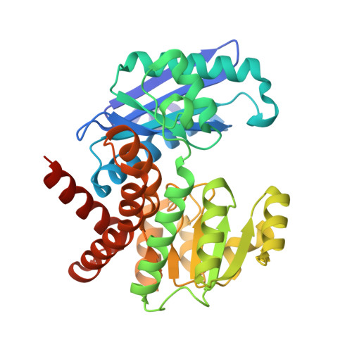
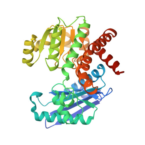
Rhodococcus L-phenylalanine dehydrogenase: kinetics, mechanism, and structural basis for catalytic specificity.

Brunhuber, N.M.Thoden, J.B.Blanchard, J.S.Vanhooke, J.L.

(2000) Biochemistry 39: 9174-9187

  • DOI: https://doi.org/10.1021/bi000494c
  • Primary Citation Related Structures: 
    1C1D, 1C1X

  • PubMed Abstract: 

    Phenylalanine dehydrogenase catalyzes the reversible, pyridine nucleotide-dependent oxidative deamination of L-phenylalanine to form phenylpyruvate and ammonia. We have characterized the steady-state kinetic behavior of the enzyme from Rhodococcus sp. M4 and determined the X-ray crystal structures of the recombinant enzyme in the complexes, E.NADH.L-phenylalanine and E.NAD(+). L-3-phenyllactate, to 1.25 and 1.4 A resolution, respectively. Initial velocity, product inhibition, and dead-end inhibition studies indicate the kinetic mechanism is ordered, with NAD(+) binding prior to phenylalanine and the products' being released in the order of ammonia, phenylpyruvate, and NADH. The enzyme shows no activity with NADPH or other 2'-phosphorylated pyridine nucleotides but has broad activity with NADH analogues. Our initial structural analyses of the E.NAD(+).phenylpyruvate and E.NAD(+). 3-phenylpropionate complexes established that Lys78 and Asp118 function as the catalytic residues in the active site [Vanhooke et al. (1999) Biochemistry 38, 2326-2339]. We have studied the ionization behavior of these residues in steady-state turnover and use these findings in conjunction with the structural data described both here and in our first report to modify our previously proposed mechanism for the enzymatic reaction. The structural characterizations also illuminate the mechanism of the redox specificity that precludes alpha-amino acid dehydrogenases from functioning as alpha-hydroxy acid dehydrogenases.


  • Organizational Affiliation
    • Department of Biochemistry, Albert Einstein College of Medicine, Bronx, New York 10461, USA.

Macromolecule Content 

  • Total Structure Weight: 74.87 kDa 
  • Atom Count: 6,119 
  • Modeled Residue Count: 697 
  • Deposited Residue Count: 710 
  • Unique protein chains: 2

Macromolecules

Find similar proteins by:|  3D Structure
Entity ID: 1
MoleculeChains  Sequence LengthOrganismDetailsImage
L-PHENYLALANINE DEHYDROGENASE355Rhodococcus sp. (in: high G+C Gram-positive bacteria)Mutation(s): 0 
EC: 1.4.1.20
UniProt
Find proteins for Q59771 (Rhodococcus sp)
Explore Q59771 
Go to UniProtKB:  Q59771
Entity Groups
Sequence Clusters30% Identity50% Identity70% Identity90% Identity95% Identity100% Identity
UniProt GroupQ59771
Sequence Annotations
Expand
Reference Sequence
Find similar proteins by:|  3D Structure
Entity ID: 2
MoleculeChains  Sequence LengthOrganismDetailsImage
L-PHENYLALANINE DEHYDROGENASE355Rhodococcus sp. (in: high G+C Gram-positive bacteria)Mutation(s): 0 
EC: 1.4.1.20
UniProt
Find proteins for Q59771 (Rhodococcus sp)
Explore Q59771 
Go to UniProtKB:  Q59771
Entity Groups
Sequence Clusters30% Identity50% Identity70% Identity90% Identity95% Identity100% Identity
UniProt GroupQ59771
Sequence Annotations
Expand
Reference Sequence

Small Molecules

Ligands 6 Unique
IDChains Name / Formula / InChI Key2D Diagram3D Interactions
NAI

Query on NAI


Download:Ideal Coordinates CCD File
F [auth A],
M [auth B]
1,4-DIHYDRONICOTINAMIDE ADENINE DINUCLEOTIDE
C21 H29 N7 O14 P2
BOPGDPNILDQYTO-NNYOXOHSSA-N
PHE

Query on PHE


Download:Ideal Coordinates CCD File
E [auth A],
L [auth B]
PHENYLALANINE
C9 H11 N O2
COLNVLDHVKWLRT-QMMMGPOBSA-N
PO4

Query on PO4


Download:Ideal Coordinates CCD File
K [auth B]PHOSPHATE ION
O4 P
NBIIXXVUZAFLBC-UHFFFAOYSA-K
IPA

Query on IPA


Download:Ideal Coordinates CCD File
N [auth B]ISOPROPYL ALCOHOL
C3 H8 O
KFZMGEQAYNKOFK-UHFFFAOYSA-N
K

Query on K


Download:Ideal Coordinates CCD File
C [auth A],
D [auth A],
G [auth B],
H [auth B]
POTASSIUM ION
K
NPYPAHLBTDXSSS-UHFFFAOYSA-N
NA

Query on NA


Download:Ideal Coordinates CCD File
I [auth B],
J [auth B]
SODIUM ION
Na
FKNQFGJONOIPTF-UHFFFAOYSA-N

Experimental Data & Validation

Experimental Data

  • Method: X-RAY DIFFRACTION
  • Resolution: 1.25 Å
  • R-Value Free:  0.238 (Depositor) 
  • R-Value Work:  0.193 (Depositor) 
  • R-Value Observed: 0.195 (Depositor) 
Space Group: P 21 21 21
Unit Cell:
Length ( Å )Angle ( ˚ )
a = 64.8α = 90
b = 110.6β = 90
c = 113.3γ = 90
Software Package:
Software NamePurpose
TNTrefinement
DENZOdata reduction
SCALEPACKdata scaling
TNTphasing

Structure Validation

View Full Validation Report


Entry History 

Deposition Data

Revision History  (Full details and data files)

  • Version 1.0: 2000-08-30
    Type: Initial release
  • Version 1.1: 2008-04-27
    Changes: Version format compliance
  • Version 1.2: 2011-07-13
    Changes: Version format compliance
  • Version 1.3: 2019-12-11
    Changes: Advisory, Derived calculations
  • Version 1.4: 2024-02-07
    Changes: Data collection, Database references, Derived calculations