1BW9 | pdb_00001bw9

PHENYLALANINE DEHYDROGENASE STRUCTURE IN TERNARY COMPLEX WITH NAD+ AND PHENYLPYRUVATE


Experimental Data Snapshot

  • Method: X-RAY DIFFRACTION
  • Resolution: 1.50 Å
  • R-Value Work: 
    0.195 (Depositor) 

wwPDB Validation 3D Report Full Report

Validation slider image for 1BW9

This is version 1.4 of the entry. See complete history

Literature

Phenylalanine dehydrogenase from Rhodococcus sp. M4: high-resolution X-ray analyses of inhibitory ternary complexes reveal key features in the oxidative deamination mechanism.

Vanhooke, J.L.Thoden, J.B.Brunhuber, N.M.Blanchard, J.S.Holden, H.M.

(1999) Biochemistry 38: 2326-2339

  • DOI: https://doi.org/10.1021/bi982244q
  • Primary Citation Related Structures: 
    1BW9, 1BXG

  • PubMed Abstract: 

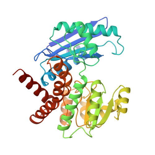
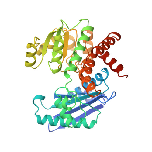
    The molecular structures of recombinant L-phenylalanine dehydrogenase from Rhodococcus sp. M4 in two different inhibitory ternary complexes have been determined by X-ray crystallographic analyses to high resolution. Both structures show that L-phenylalanine dehydrogenase is a homodimeric enzyme with each monomer composed of distinct globular N- and C-terminal domains separated by a deep cleft containing the active site. The N-terminal domain binds the amino acid substrate and contributes to the interactions at the subunit:subunit interface. The C-terminal domain contains a typical Rossmann fold and orients the dinucleotide. The dimer has overall dimensions of approximately 82 A x 75 A x 75 A, with roughly 50 A separating the two active sites. The structures described here, namely the enzyme.NAD+.phenylpyruvate, and enzyme. NAD+.beta-phenylpropionate species, represent the first models for any amino acid dehydrogenase in a ternary complex. By analysis of the active-site interactions in these models, along with the currently available kinetic data, a detailed chemical mechanism has been proposed. This mechanism differs from those proposed to date in that it accounts for the inability of the amino acid dehydrogenases, in general, to function as hydroxy acid dehydrogenases.


  • Organizational Affiliation
    • Department of Biochemistry, University of Wisconsin-Madison 53705, USA.

Macromolecule Content 

  • Total Structure Weight: 75.48 kDa 
  • Atom Count: 6,225 
  • Modeled Residue Count: 697 
  • Deposited Residue Count: 712 
  • Unique protein chains: 2

Macromolecules

Find similar proteins by:|  3D Structure
Entity ID: 1
MoleculeChains  Sequence LengthOrganismDetailsImage
PHENYLALANINE DEHYDROGENASE356Rhodococcus sp. (in: high G+C Gram-positive bacteria)Mutation(s): 0 
EC: 1.4.1.20
UniProt
Find proteins for Q59771 (Rhodococcus sp)
Explore Q59771 
Go to UniProtKB:  Q59771
Entity Groups
Sequence Clusters30% Identity50% Identity70% Identity90% Identity95% Identity100% Identity
UniProt GroupQ59771
Sequence Annotations
Expand
Reference Sequence
Find similar proteins by:|  3D Structure
Entity ID: 2
MoleculeChains  Sequence LengthOrganismDetailsImage
PHENYLALANINE DEHYDROGENASE356Rhodococcus sp. (in: high G+C Gram-positive bacteria)Mutation(s): 0 
EC: 1.4.1.20
UniProt
Find proteins for Q59771 (Rhodococcus sp)
Explore Q59771 
Go to UniProtKB:  Q59771
Entity Groups
Sequence Clusters30% Identity50% Identity70% Identity90% Identity95% Identity100% Identity
UniProt GroupQ59771
Sequence Annotations
Expand
Reference Sequence

Small Molecules

Ligands 7 Unique
IDChains Name / Formula / InChI Key2D Diagram3D Interactions
NAD

Query on NAD


Download:Ideal Coordinates CCD File
C [auth A],
J [auth B]
NICOTINAMIDE-ADENINE-DINUCLEOTIDE
C21 H27 N7 O14 P2
BAWFJGJZGIEFAR-NNYOXOHSSA-N
PPY

Query on PPY


Download:Ideal Coordinates CCD File
D [auth A],
K [auth B]
3-PHENYLPYRUVIC ACID
C9 H8 O3
BTNMPGBKDVTSJY-UHFFFAOYSA-N
PO4

Query on PO4


Download:Ideal Coordinates CCD File
I [auth B]PHOSPHATE ION
O4 P
NBIIXXVUZAFLBC-UHFFFAOYSA-K
EDO

Query on EDO


Download:Ideal Coordinates CCD File
L [auth B],
M [auth B],
N [auth B]
1,2-ETHANEDIOL
C2 H6 O2
LYCAIKOWRPUZTN-UHFFFAOYSA-N
IPA

Query on IPA


Download:Ideal Coordinates CCD File
O [auth B],
P [auth B],
Q [auth B],
R [auth B]
ISOPROPYL ALCOHOL
C3 H8 O
KFZMGEQAYNKOFK-UHFFFAOYSA-N
K

Query on K


Download:Ideal Coordinates CCD File
E [auth B],
H [auth B]
POTASSIUM ION
K
NPYPAHLBTDXSSS-UHFFFAOYSA-N
NA

Query on NA


Download:Ideal Coordinates CCD File
F [auth B],
G [auth B]
SODIUM ION
Na
FKNQFGJONOIPTF-UHFFFAOYSA-N

Experimental Data & Validation

Experimental Data

  • Method: X-RAY DIFFRACTION
  • Resolution: 1.50 Å
  • R-Value Work:  0.195 (Depositor) 
Space Group: P 21 21 21
Unit Cell:
Length ( Å )Angle ( ˚ )
a = 64.3α = 90
b = 110.2β = 90
c = 113.4γ = 90
Software Package:
Software NamePurpose
TNTrefinement
SAINTdata reduction
XCALIBREdata scaling

Structure Validation

View Full Validation Report


Entry History 

Deposition Data

Revision History  (Full details and data files)

  • Version 1.0: 1999-05-18
    Type: Initial release
  • Version 1.1: 2008-03-25
    Changes: Version format compliance
  • Version 1.2: 2011-07-13
    Changes: Version format compliance
  • Version 1.3: 2019-12-11
    Changes: Advisory, Derived calculations, Other
  • Version 1.4: 2024-02-07
    Changes: Data collection, Database references, Derived calculations