1BVX | pdb_00001bvx

THE 1.8 A STRUCTURE OF GEL GROWN TETRAGONAL HEN EGG WHITE LYSOZYME


Experimental Data Snapshot

  • Method: X-RAY DIFFRACTION
  • Resolution: 1.80 Å
  • R-Value Free: 
    0.217 (Depositor) 
  • R-Value Work: 
    0.182 (Depositor), 0.190 (DCC) 
  • R-Value Observed: 
    0.182 (Depositor) 

Starting Model: experimental
View more details

wwPDB Validation 3D Report Full Report

Validation slider image for 1BVX

This is version 1.4 of the entry. See complete history

Literature

Bound-solvent structures for microgravity-, ground control-, gel- and microbatch-grown hen egg-white lysozyme crystals at 1.8 A resolution.

Dong, J.Boggon, T.J.Chayen, N.E.Raftery, J.Bi, R.C.Helliwell, J.R.

(1999) Acta Crystallogr D Biol Crystallogr 55: 745-752

  • DOI: https://doi.org/10.1107/s0907444998016047
  • Primary Citation Related Structures: 
    1BVX, 1BWH, 1BWI, 1BWJ

  • PubMed Abstract: 

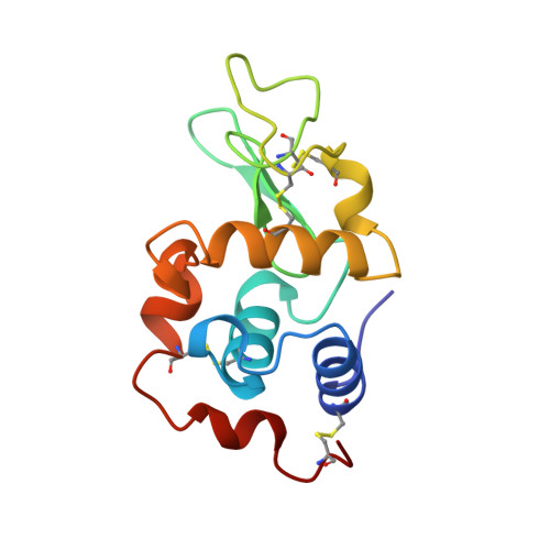
    A number of methods can be used to improve the stability of the protein crystal-growth environment, including growth in microgravity without an air-liquid phase boundary, growth in gels and growth under oil ('microbatch'). In this study, X-ray data has been collected from and structures refined for crystals of hen egg-white lysozyme (HEWL) grown using four different methods, liquid-liquid dialysis on Earth and in microgravity using the European Space Agency's (ESA) Advanced Protein Crystallization Facility (APCF) on board the NASA Space Shuttle Life and Microgravity Spacelab (LMS) mission (STS-78), crystallization in agarose gel using a tube liquid-gel diffusion method and crystallization in microbatch under oil. A comparison of the overall quality of the X-ray data, the protein structures and especially the bound-water structures has been carried out at 1.8 A. The lysozyme protein structures corresponding to these four different crystallization methods remain similar. A small improvement in the bound-solvent structure is seen in lysozyme crystals grown in microgravity by liquid-liquid dialysis, which has a more stable fluid physics state in microgravity, and is consistent with a better formed protein crystal in microgravity.


  • Organizational Affiliation
    • Section of Structural Chemistry, Department of Chemistry, University of Manchester M13 9PL, England.

Macromolecule Content 

  • Total Structure Weight: 14.33 kDa 
  • Atom Count: 1,096 
  • Modeled Residue Count: 129 
  • Deposited Residue Count: 129 
  • Unique protein chains: 1

Macromolecules

Find similar proteins by:|  3D Structure
Entity ID: 1
MoleculeChains  Sequence LengthOrganismDetailsImage
PROTEIN (LYSOZYME)129Gallus gallusMutation(s): 0 
EC: 3.2.1.17
UniProt
Find proteins for P00698 (Gallus gallus)
Explore P00698 
Go to UniProtKB:  P00698
Entity Groups
Sequence Clusters30% Identity50% Identity70% Identity90% Identity95% Identity100% Identity
UniProt GroupP00698
Sequence Annotations
Expand
Reference Sequence

Experimental Data & Validation

Experimental Data

  • Method: X-RAY DIFFRACTION
  • Resolution: 1.80 Å
  • R-Value Free:  0.217 (Depositor) 
  • R-Value Work:  0.182 (Depositor), 0.190 (DCC) 
  • R-Value Observed: 0.182 (Depositor) 
Space Group: P 43 21 2
Unit Cell:
Length ( Å )Angle ( ˚ )
a = 79.203α = 90
b = 79.203β = 90
c = 38.04γ = 90
Software Package:
Software NamePurpose
DENZOdata reduction
SCALEPACKdata scaling
X-PLORrefinement

Structure Validation

View Full Validation Report


Entry History 

Deposition Data

Revision History  (Full details and data files)

  • Version 1.0: 1998-09-23
    Type: Initial release
  • Version 1.1: 2008-04-27
    Changes: Version format compliance
  • Version 1.2: 2011-07-13
    Changes: Version format compliance
  • Version 1.3: 2023-08-09
    Changes: Data collection, Database references, Refinement description
  • Version 1.4: 2024-10-30
    Changes: Structure summary