1BVK | pdb_00001bvk

HUMANIZED ANTI-LYSOZYME FV COMPLEXED WITH LYSOZYME


Experimental Data Snapshot

  • Method: X-RAY DIFFRACTION
  • Resolution: 2.70 Å
  • R-Value Free: 
    0.297 (Depositor) 
  • R-Value Work: 
    0.208 (Depositor) 
  • R-Value Observed: 
    0.208 (Depositor) 

Starting Model: experimental
View more details

wwPDB Validation 3D Report Full Report

Validation slider image for 1BVK

This is version 1.4 of the entry. See complete history

Literature

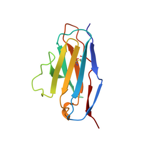
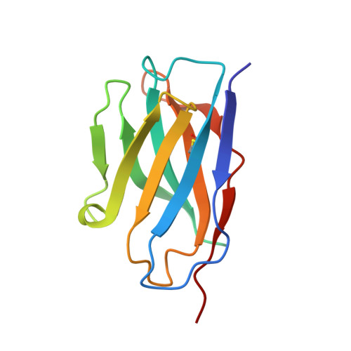
Conformational correction mechanisms aiding antigen recognition by a humanized antibody.

Holmes, M.A.Buss, T.N.Foote, J.

(1998) J Exp Medicine 187: 479-485

  • DOI: https://doi.org/10.1084/jem.187.4.479
  • Primary Citation Related Structures: 
    1BVK

  • PubMed Abstract: 

    The crystal structure of the complex between hen egg lysozyme and the Fv fragment of a humanized antilysozyme antibody was determined to 2.7-A resolution. The structure of the antigen combining site in the complex is nearly identical to that of the complexed form of the parent mouse antibody, D1.3. In contrast, the combining sites of the unliganded mouse and humanized antilysozymes show moderate conformational differences. This disparity suggests that a conformational readjustment process linked to antigen binding reverses adverse conformations in the complementarity determining regions that had been introduced by engineering these segments next to human framework regions in the humanized antibody.


  • Organizational Affiliation
    • Division of Molecular Medicine, Fred Hutchinson Cancer Research Center, Seattle, Washington 98109-1024, USA.

Macromolecule Content 

  • Total Structure Weight: 78.33 kDa 
  • Atom Count: 5,488 
  • Modeled Residue Count: 706 
  • Deposited Residue Count: 708 
  • Unique protein chains: 3

Macromolecules

Find similar proteins by:|  3D Structure
Entity ID: 1
MoleculeChains  Sequence LengthOrganismDetailsImage
HULYS11
A, D
108Homo sapiensMutation(s): 0 
Find similar proteins by:|  3D Structure
Entity ID: 2
MoleculeChains  Sequence LengthOrganismDetailsImage
HULYS11
B, E
117Homo sapiensMutation(s): 0 
Find similar proteins by:|  3D Structure
Entity ID: 3
MoleculeChains  Sequence LengthOrganismDetailsImage
LYSOZYME
C, F
129Gallus gallusMutation(s): 0 
EC: 3.2.1.17
UniProt
Find proteins for P00698 (Gallus gallus)
Explore P00698 
Go to UniProtKB:  P00698
Entity Groups
Sequence Clusters30% Identity50% Identity70% Identity90% Identity95% Identity100% Identity
UniProt GroupP00698
Sequence Annotations
Expand
Reference Sequence

Experimental Data & Validation

Experimental Data

  • Method: X-RAY DIFFRACTION
  • Resolution: 2.70 Å
  • R-Value Free:  0.297 (Depositor) 
  • R-Value Work:  0.208 (Depositor) 
  • R-Value Observed: 0.208 (Depositor) 
Space Group: P 41 21 2
Unit Cell:
Length ( Å )Angle ( ˚ )
a = 97.7α = 90
b = 97.7β = 90
c = 174.9γ = 90
Software Package:
Software NamePurpose
DENZOdata reduction
SCALEPACKdata scaling
X-PLORmodel building
X-PLORrefinement
X-PLORphasing

Structure Validation

View Full Validation Report


Entry History 

Deposition Data

Revision History  (Full details and data files)

  • Version 1.0: 1999-02-16
    Type: Initial release
  • Version 1.1: 2008-03-24
    Changes: Version format compliance
  • Version 1.2: 2011-07-13
    Changes: Version format compliance
  • Version 1.3: 2023-08-09
    Changes: Database references, Refinement description
  • Version 1.4: 2024-10-23
    Changes: Data collection, Structure summary